




İçeriğe geç
Kudret Eye Istanbul

Our Laser Technology

  • Last updated:

Laser vision correction has a proven record of safety and predictability since the 1990s; what decides the outcome is the quality of the equipment and the accuracy of the measurements. The laser and diagnostic devices we use at Kudret Eye, and what each one does, are below.

Examination and imaging devices

Johnson & Johnson Vision

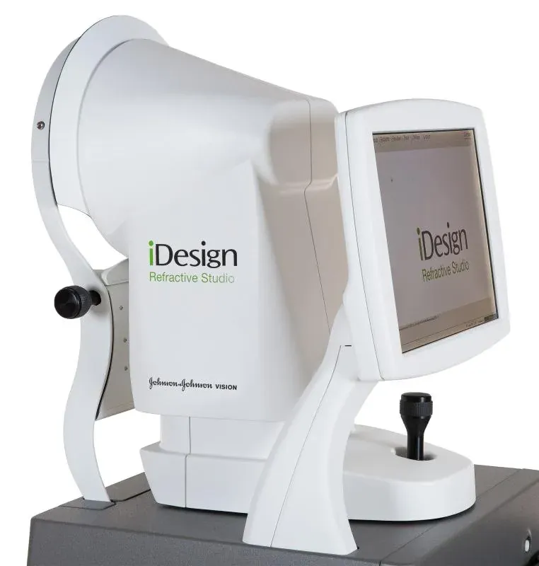
iDesign Advanced WaveScan Studio

The measurement device that captures the eye’s optical “fingerprint”. In a single scan it records refractive error at over 1,200 points, corneal topography and pupil metrics. The map is passed to the excimer laser, so treatment follows your eye’s own map rather than a standard prescription.

Why we chose this device

A spectacle prescription is a coarse summary of the eye's refractive error; this device takes thousands of separate readings across the same eye and maps them. Laser treatment is then planned around that eye's own pattern rather than an average figure, which is where night vision is won or lost.
Manufacturer's page

iDesign Advanced WaveScan Studio

Tracey Technologies

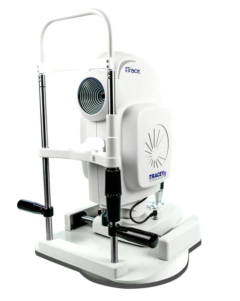
iTrace

A vision-quality analyser that splits the eye’s total refractive error into the parts coming from the cornea and from the natural lens. One of the measurements the surgeon relies on when deciding between laser and lens replacement, and when choosing a trifocal lens.

Why we chose this device

One of the methods that can tell whether a visual complaint comes from the cornea or from the lens inside the eye. That distinction changes the treatment: a corneal cause calls for laser, a lens cause for lens exchange. By ray-tracing the two separately, it keeps patients from being steered to the wrong procedure.

Manufacturer's page

iTrace

Laser systems

ZEISS VisuMax 800

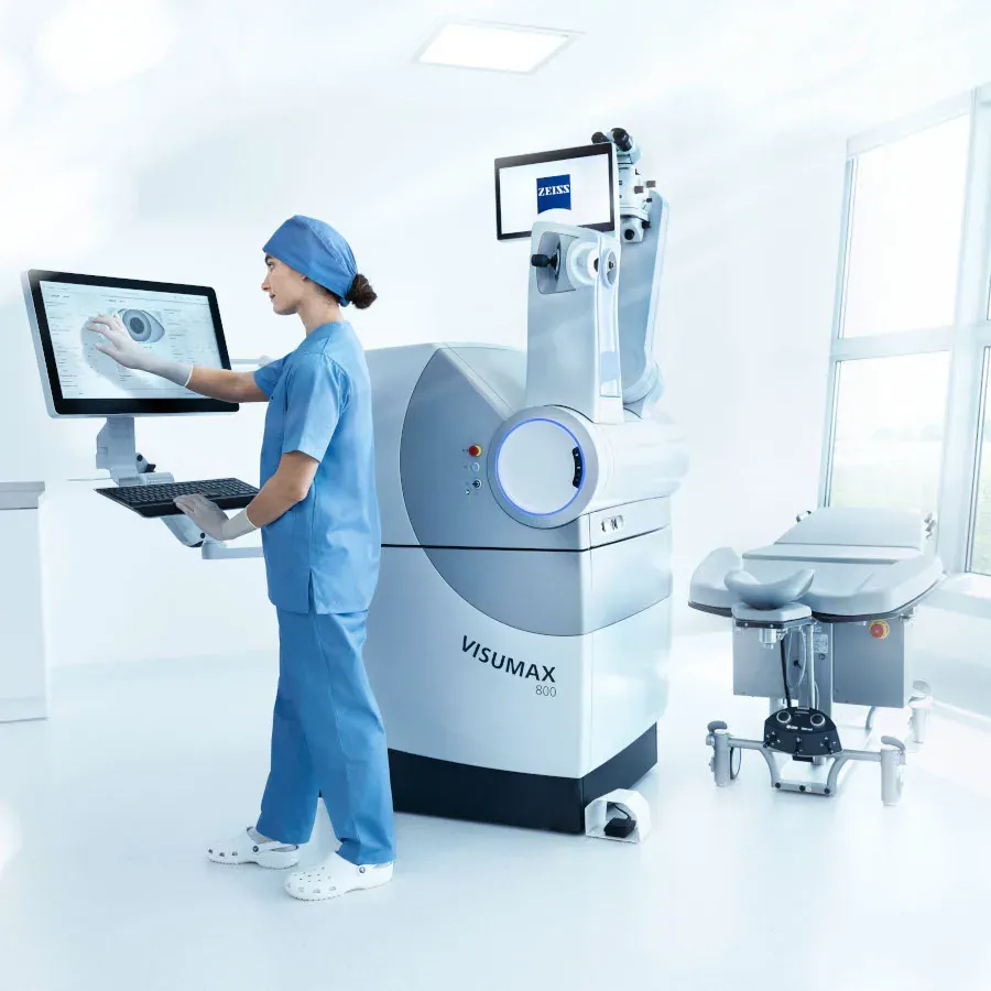
The femtosecond laser behind ReLEx SMILE PRO. It creates a thin lenticule inside the cornea and removes it through a 2-4 mm incision; no flap is cut. Lenticule cutting takes under 10 seconds per eye and the device tracks eye movement to centre itself.

Why we chose this device

On this laser the lenticule cut in SMILE takes seconds; the shorter the eye has to be held still, the smaller the margin for error from patient movement. Its eye tracking keeps the cut on the planned axis even if the eye shifts.

Manufacturer's page

ZEISS VisuMax 800

ZEISS VISUMAX 500

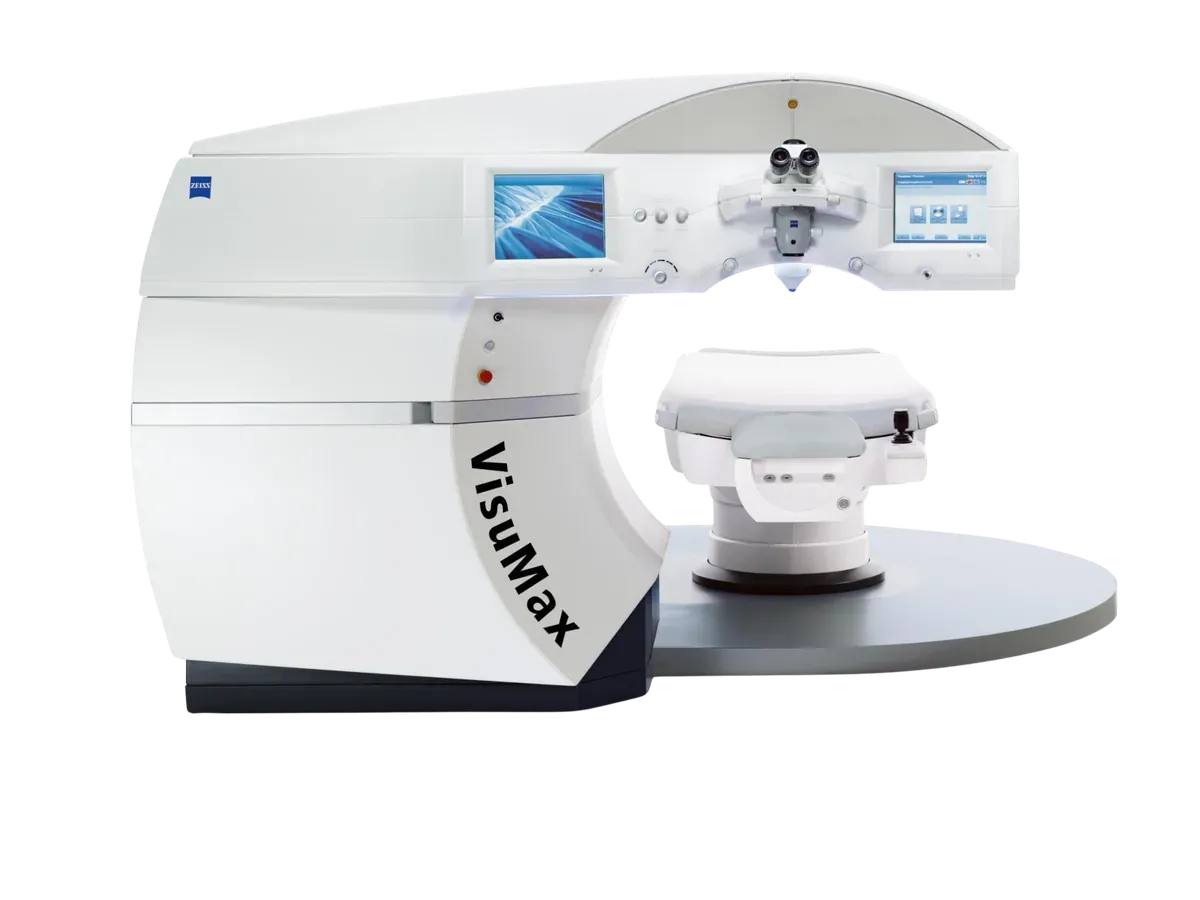
The femtosecond laser the ReLEx SMILE technique was developed on. It shapes a thin lens (lenticule) inside the cornea and removes it through a small incision, without raising a flap on the surface. A suction ring holds the eye with gentle contact, and the curved interface, matched to the eye's own curvature, spreads the pressure.

Why we chose this device

Most of the SMILE literature rests on results gathered on this platform, so the long-term data for the technique comes from this very device. Having it alongside the VISUMAX 800 means two femtosecond lasers: we can run two rooms on the same day, and a service visit does not push the surgery schedule.

Manufacturer's page

ZEISS VISUMAX 500

Johnson & Johnson Vision

IntraLase iFS 150 kHz

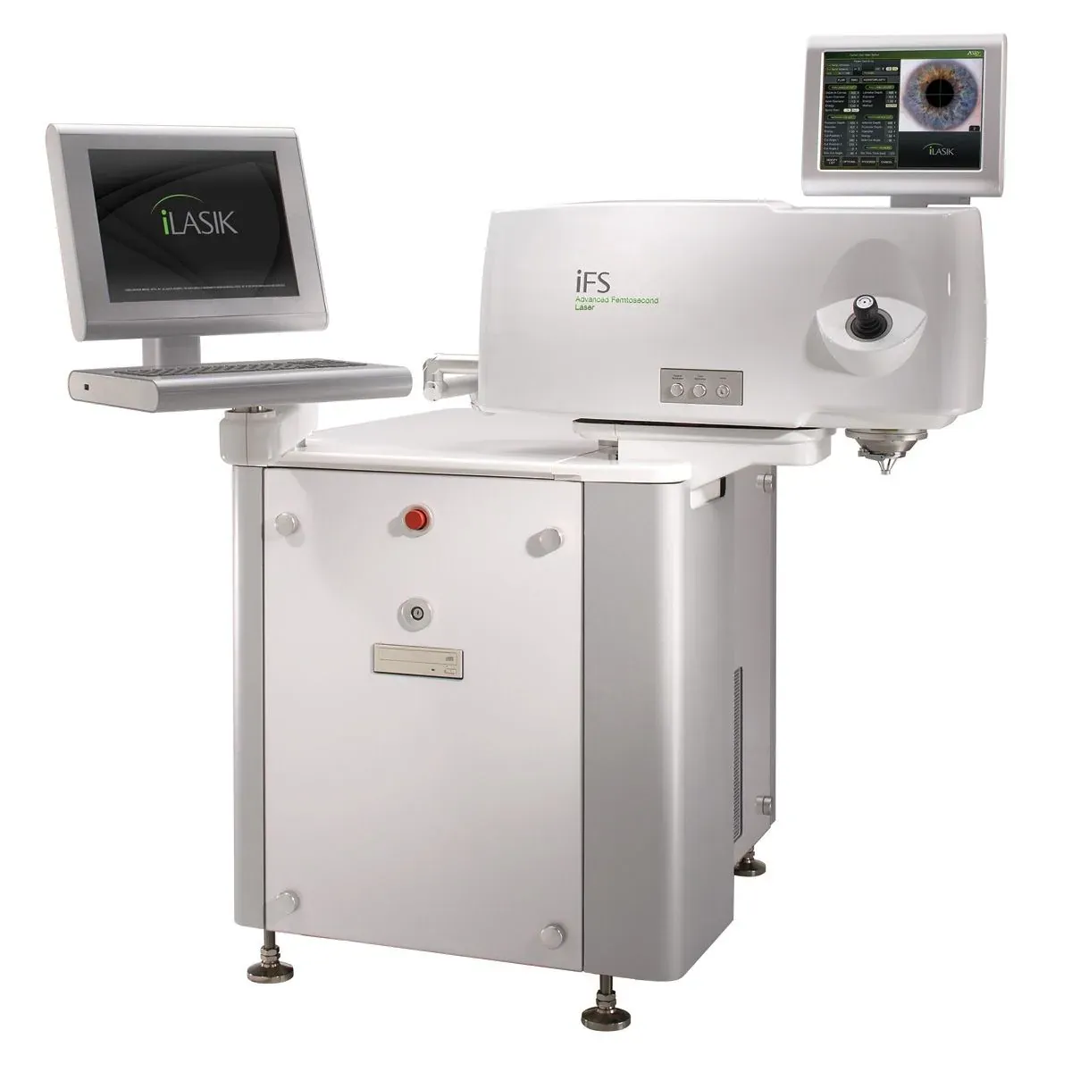
The first step of iLASIK: the corneal flap is created 100 % blade-free with a femtosecond laser instead of a blade. At 150 kHz the flap forms in seconds, at the exact thickness and shape the surgeon sets, far more predictable than the older microkeratome. The device is the first link in the iLASIK platform developed by Abbott Medical Optics, today part of Johnson & Johnson Vision.

Why we chose this device

Creating the flap with a laser rather than a blade keeps its thickness uniform to the micron across the whole cornea. The 150 kHz speed shortens the step, and the bevelled edge lets the flap seat back more securely.

Manufacturer's page

IntraLase iFS 150 kHz

Ziemer

FEMTO LDV Z8

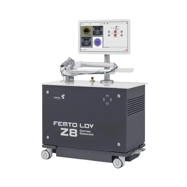
A mobile femtosecond laser usable in both corneal and cataract surgery. Its pulse energy is very low and its repetition rate very high, so cuts are formed from small overlapping spots. It can raise a flap, and in cataract surgery it opens the front capsule of the lens and fragments the lens itself. Being on wheels, the laser comes to the table rather than the patient being moved.

Why we chose this device

Opening the front capsule by hand is one of the steps in cataract surgery that most depends on the surgeon's experience; a laser-made capsulotomy comes out the same diameter and centred in every patient, which helps the intraocular lens sit where it should. That the device can be brought to the patient matters too: nobody is moved from one table to another mid-procedure.

Manufacturer's page

FEMTO LDV Z8

Johnson & Johnson Vision

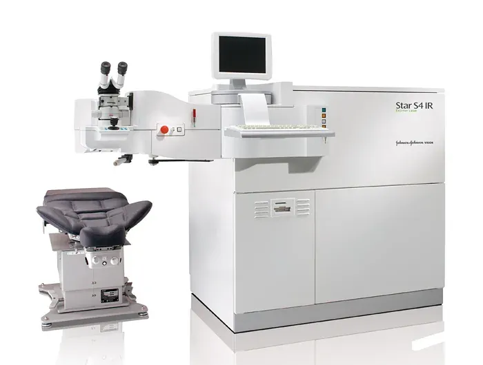
VISX STAR S4 IR Excimer

The excimer laser of iLASIK’s second step. It reshapes corneal tissue with micron precision; infrared eye tracking keeps every pulse on target even if the eye moves. Paired with iDesign measurements it delivers a fully personalised (wavefront-guided) treatment. This laser is part of the iLASIK platform developed by Abbott Medical Optics, today part of Johnson & Johnson Vision.

Why we chose this device

Infrared eye tracking measures the small involuntary movements of the eye hundreds of times a second and keeps the laser on the same spot. Its variable spot size removes only as much tissue as needed, and the less cornea spent now, the more margin remains for a later touch-up.

Manufacturer's page

VISX STAR S4 IR Excimer

We keep renewing our equipment

Devices in eye care change quickly, and a newer system often means a shorter, more comfortable or more precise examination. We have renewed our equipment regularly since the day we opened, bringing new-generation systems into the clinic. This page is not fixed either: when a new device arrives, it is added here.

The laser evaluation process

Whether you are suitable for laser is decided in a single examination with the measurements below. It takes about an hour and a half; contact-lens wearers are asked to leave soft lenses out for 5 days and hard lenses for 2 weeks beforehand.

  1. Initial consultation

    Your expectations, medical history and glasses or lens use are discussed.

  2. Visual acuity

    The vision of both eyes is measured with and without correction.

  3. Refraction

    Your prescription is confirmed both by machine and by the surgeon.

  4. Corneal topography

    A surface map of the cornea is taken; conditions such as keratoconus show up here.

  5. Corneal thickness

    Pachymetry measures the thickness of the cornea, which sets the safe limit for laser.

  6. Pupil measurement

    Pupil size is measured in dim light and feeds into night-vision planning.

  7. Dry-eye test

    Tear volume and quality are assessed; dryness is treated first if present.

  8. Retinal examination

    With the pupil dilated, the retina and optic nerve are checked.

  9. Results and choice of method

    Your surgeon reviews all the measurements and recommends the most suitable method.

FREE CONSULTATION

Your appointment request has been received successfully.

We will be in touch with you as soon as possible.

If your details are not correct, please send a new request.

Your cookie choice

Cookies needed for the site to work are always on. With your permission we also use Google and Meta cookies to measure visits and show you relevant ads. You can change your choice at any time with the "Cookie settings" link at the bottom of the page.Detailed Information

Suitability test

Which treatment suits me?

A few short questions to show you which treatments would be discussed at your examination. At the end we will call you for a free consultation.

Book a Free Consultation

FREE CONSULTATION

Site search

Can't find what you're looking for?

We are just a phone call away.