


 Türkçe
 English
 Deutsch
 Nederlands
 Italiano
 Español
 Français
 Български
 Română
 Русский
 العربية


Kudret Eye IstanbulRetina
Attention to Floaters in Front of the Eyes!
Kudret Eye İstanbul Chief Physician Op. Dr. Nail Sönmez warns that floaters in front of the eye may come from natural changes in the eye or be an…

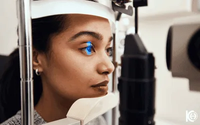
Retinal treatments are various methods used to treat retinal diseases. These include injections, laser therapy and surgical interventions; their aim is to maintain retinal health and prevent vision loss.
Vitrectomy Surgery is a surgical operation applied for diseases occurring in the back of the eye. Bleeding may occur in the eye for a number of reasons. At the same time, membrane formation may occur in the eye due to diabetes. Vitrectomy surgery is needed both to clear the blood from the eye and to peel the membranes formed in the eye.
MACROvision is an intraocular optical implant used in retinal diseases that impair central vision, such as macular degeneration. It magnifies the image and directs it to the healthy part of the macula; it does not treat the underlying disease, but helps the patient make better use of the vision that remains.
Macula Degeneration is characterised by damage to the central area of the retina. This condition affects central vision and usually occurs with age. The disease is divided into two types: dry type and wet type. The dry type is caused by the deterioration of cells in the macular layer, while the wet type is characterised by the formation of new, abnormal vessels, which can lead to leakage and loss of vision.
Eye health is an important part of general health and retinal diseases are serious conditions that threaten eye health. The retina is a sensitive tissue located at the back of the eye that detects light and transmits the image to the brain. Retinal diseases can cause vision loss by impairing the function of this critical tissue. Early diagnosis and effective treatment play an important role in the management of retinal diseases.
Retinal detachment, an eye problem that seriously threatens vision, is the separation of the retinal nerve layer from the pigment epithelium layer underneath. It usually develops when fluid leaks between these two layers following a tear or hole in the retina. This separation is similar to the separation of a damp wallpaper from the wall.
Diabetic Retinopathy is a complication that occurs in diabetic patients and affects the retina, the light sensitive layer at the back of the eye. High blood sugar levels can damage the capillaries in the retina, leading to vision problems and blindness in progressive cases.
Retinopathy of Prematurity (ROP) is an eye disease that occurs in premature and low birth weight babies with abnormal growth of blood vessels in the retina, the light-sensitive tissue layer behind the eye.
Over 24 years in eye care: three eye hospitals and a general hospital, a team of specialist surgeons and technology renewed every year.
Hundreds of thousands of successful operations and a name trusted across generations.
World-leading equipment for SMILE PRO, iLASIK and trifocal lenses.
Every treatment is performed by a surgeon who specialises in it.
Follow-up checks and, if ever needed, enhancement are our responsibility.
They made the process of coming to a country where I don’t speak the language and having eye surgery (frightening enough in your home town) very easy indeed. I had a wonderful interpreter with me through the entire process every day, she made everything so easy. First class facilities, first class staff and a very convenient location.

A very kind and professional medical team! The surgery was completely straightforward and was completed relatively quickly. The entire team, and especially Zuhal, my patient coordinator, was always available, incredibly helpful, and genuinely pleasant throughout the whole process. She stayed by my side from start to finish, explained everything thoroughly, and always made sure I understood what was happening. The doctor worked in a very calm and professional manner, and I never felt uncertain or uncomfortable at any point. There were three fresh meals each day: breakfast, lunch, and dinner, and they were all very enjoyable. The hospital rooms were also very clean and well maintained. Overall, I had a very positive experience with this hospital. I’m truly grateful to the entire team and would wholeheartedly recommend this hospital to anyone.
Easy process to book appointment, clear communication throughout. The hospital I clean and well-equipped. The staff friendly and helpful. A translator on hand from the moment I arrived until I left The preparation and tests pror to the Yag laser procedure, faultless. Clear information given on each part if the process. The procedure was painless, and no ill effects and I could see clearly about 30 minutes after the procedure. I cannot recommend them enough .
Operation Duration
30 minutes to 2 hours.
Recovery Time
Back to daily life in a few days, full recovery in 2-4 weeks
Anaesthesia Type
Local or General Anaesthesia
Kudret Eye İstanbul Chief Physician Op. Dr. Nail Sönmez warns that floaters in front of the eye may come from natural changes in the eye or be an…
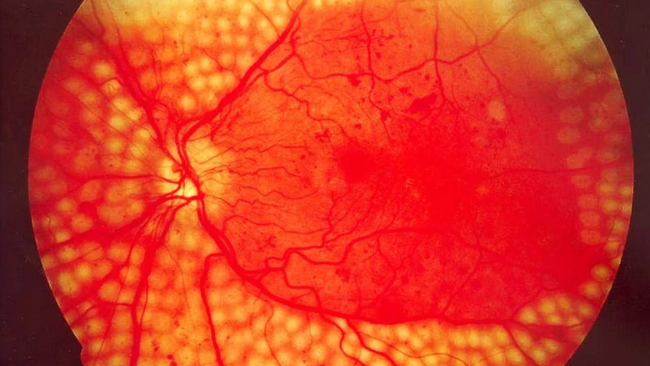
Diabetic retinopathy is the greatest cause of sight loss arising from diabetes. Raised blood sugar damages the blood vessels, and diabetes affects…
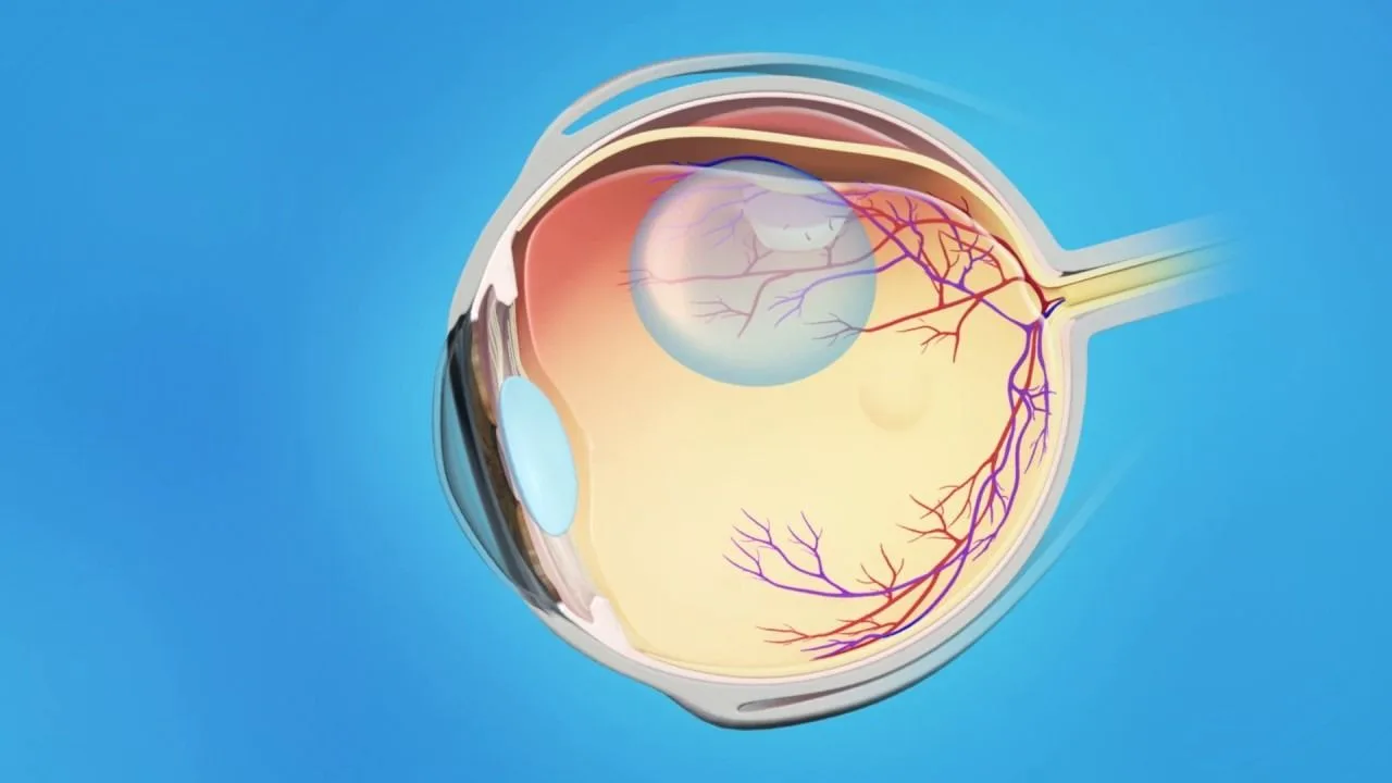
Retinal detachment occurs as fluid gathers beneath the light-sensitive retinal layer and the sensory retina separates from the pigment epithelium. It…
Your cookie choice
Essential cookies are always on. With your permission we also use Google and Meta cookies for measurement and ads.Cookies needed for the site to work are always on. With your permission we also use Google and Meta cookies to measure visits and show you relevant ads. You can change your choice at any time with the "Cookie settings" link at the bottom of the page.Detailed Information