Eye Examination
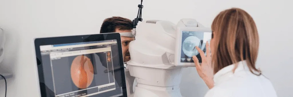
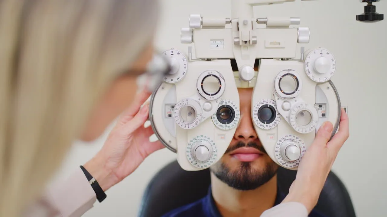
Eye examination is a medical check-up that is recommended to be performed at regular intervals and is important for maintaining eye health and diagnosing various vision problems at an early stage. During this examination, the retina and general eye health, especially eye pressure, are evaluated in detail. During the eye examination, various tests are used to measure vision and examine the different functions of the eye. These examinations carried out by the ophthalmologist cover a wide range of aspects, from visual acuity and colour perception to the function of the eye muscles and eye pressure. In this way, symptoms of any visual impairment or eye disease can be detected early and necessary interventions can be made in a timely manner. 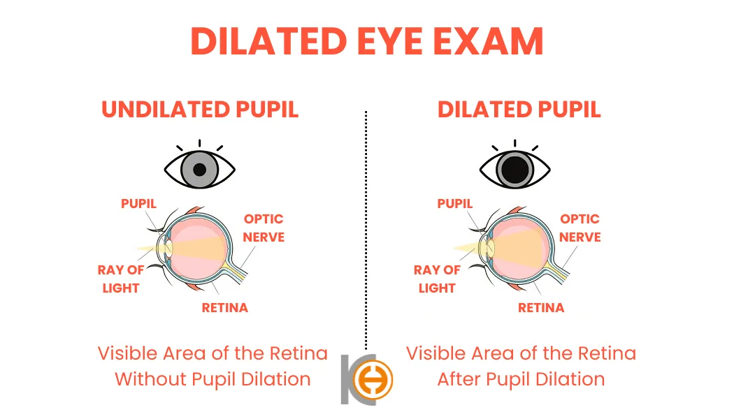
Diagnosis of Refractive Defects
Diagnosis of refractive errors aims to identify conditions in which the eye cannot focus light properly. Refractive errors can lead to problems such as poor near or far vision, headaches and eye fatigue. Fortunately, these defects can be detected with a simple eye examination and then corrected by various methods such as glasses, contact lenses or laser surgery. In this way, the quality of life of patients improves and it is possible to have a clearer vision.
Colour Blindness Test
The colour blindness test is an assessment method designed to detect deficiencies or impairments in the ability to perceive colour. This disorder, which is often detected in childhood, is manifested by difficulty in perceiving certain colours or tones. Colour blindness, which can affect daily life in many ways, can be easily diagnosed with various tests and can be managed with different strategies according to the individual's condition.
Which tests are done at an eye examination?
An eye examination is not a single measurement but a series of tests that build on one another. Each looks at a different layer of the eye, and the doctor reads them together. Below is what is done, in order, and why each step is needed.
Visual acuity
The examination starts by reading letters from a chart. This is the only measurement that shows how the eye is performing right now. Your prescription may have changed, but reduced vision can just as easily come from the cornea or the retina. When acuity is down, the remaining tests say where to look.
Refraction
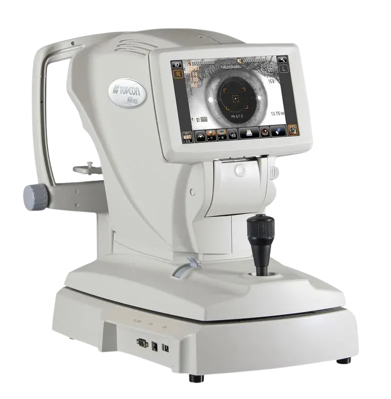
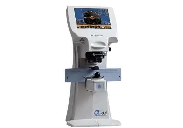
Your myopia, hyperopia and astigmatism are measured automatically first. That reading is not the prescription itself but the starting point: the doctor works from it towards the value at which you see most clearly. The prescription of the glasses you already wear is read on a separate device, so it is clear whether anything has actually changed.
Eye pressure
Raised eye pressure is the one finding that can quietly damage the optic nerve over years while causing no symptoms at all. Caught early, glaucoma can be halted; caught late, the vision that has gone does not come back. That is why pressure is measured even when there is no complaint. A non-contact tonometer does this with a short puff of air, without touching the eye and without drops.
Slit lamp examination
The doctor examines the front half of the eye under magnification: the eyelids, the cornea, the anterior chamber and the lens. Cataract, dry eye, corneal clouding and inflammation are seen here.
Corneal mapping
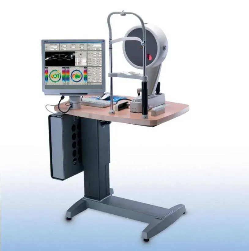
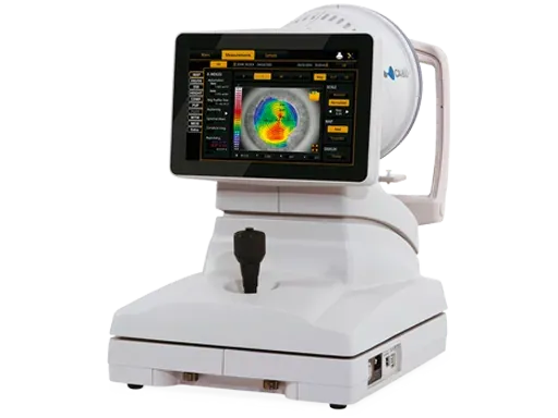
The surface of the cornea is mapped: its curvature, its thickness and how regular it is. This answers two questions at once. If the astigmatism is irregular, the map shows that glasses will not correct it. And if laser treatment is being considered, the map shows whether the cornea allows it. A progressive thinning such as keratoconus also shows up here first, before the patient notices anything.
Tear film and eyelid assessment
How long the tear film holds and whether the oil glands in the eyelid are working are both measured. Burning, stinging and vision that fluctuates through the day often come from this rather than from a change in prescription. It matters before laser surgery too: a dry eye needs treating first.
Dilated examination of the back of the eye
Where it is needed, drops widen the pupil so the back half of the eye, the retina and the optic nerve, can be assessed. Retinal tears, macular disease and the marks diabetes leaves in the eye are found at this stage. For a few hours afterwards near vision is blurred and the eye is sensitive to light, so you should not drive during that time.
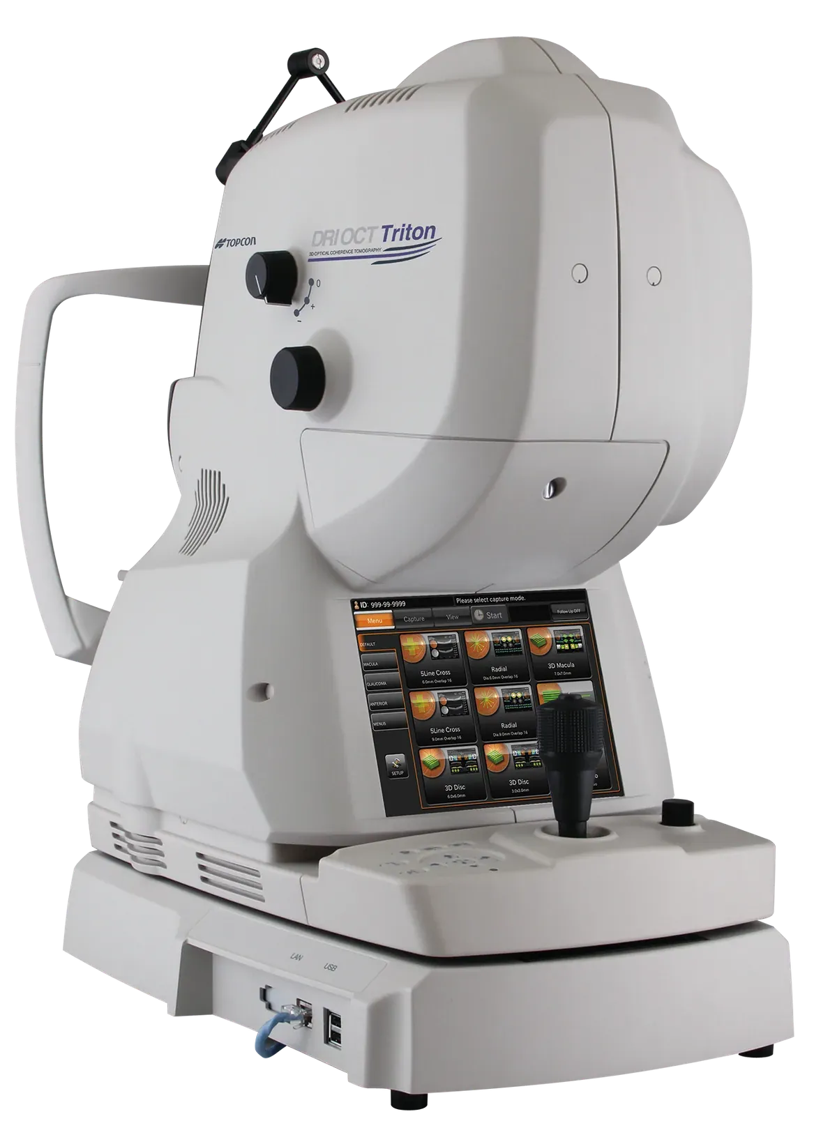
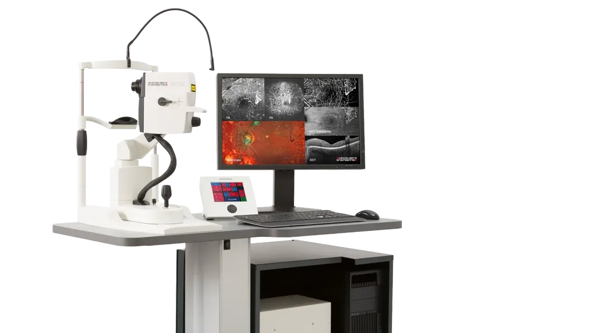
Retinal imaging (OCT)
The layers of the retina are imaged as cross sections at micron level. Fluid, thinning and swelling that cannot be seen by looking at the back of the eye appear here. In macular disease and in diabetic changes, repeating the same scan is also how it is judged whether treatment is working.
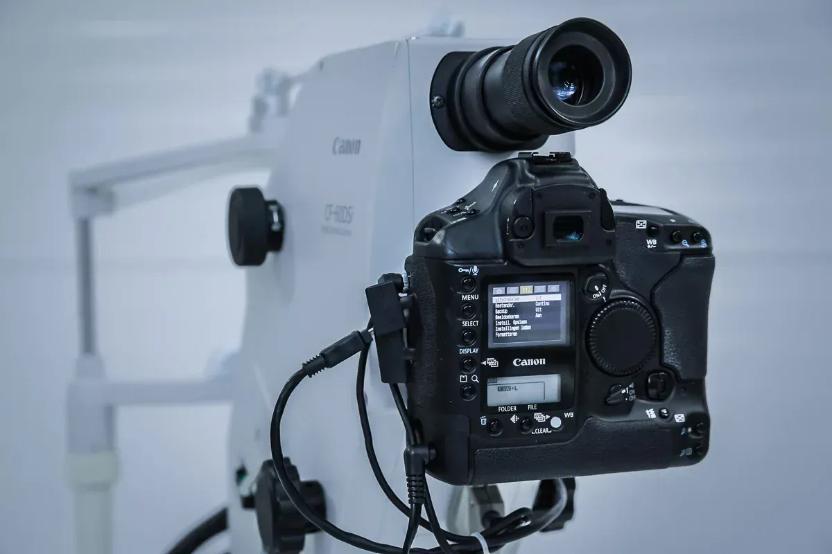
Fundus photography
A colour photograph is taken of the retina, the macula and the optic nerve. On its own it may say little; its value is in comparison. Put side by side with the photograph taken a year later, any change is beyond argument.
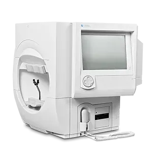
Visual field
Peripheral vision can be lost while central vision stays sharp, and people often do not notice for a long time. The visual field test maps where the loss is. It is the cornerstone of diagnosing and following glaucoma, because it shows what the damage to the optic nerve actually costs in function.
Corneal cell count
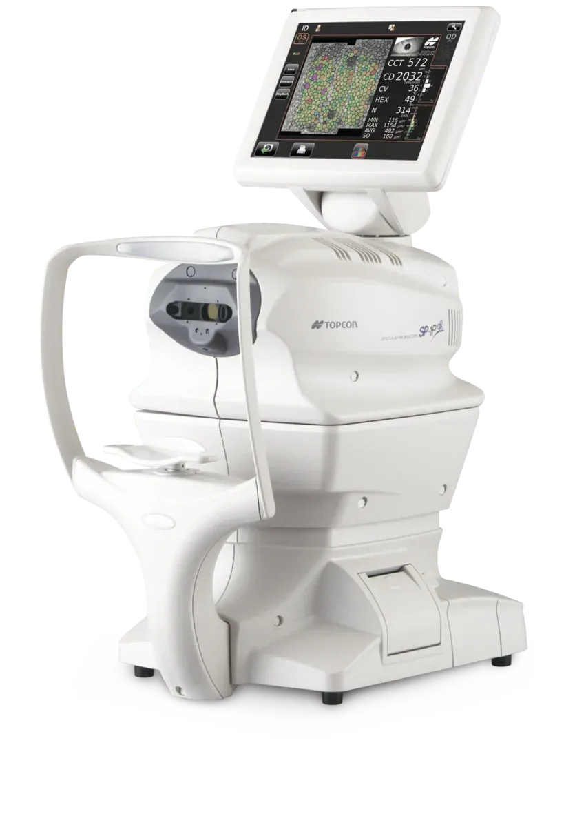
The cells in the innermost layer of the cornea keep it clear, and they do not regenerate. Their number says in advance how much surgery an eye can tolerate. In eyes being considered for cataract or lens surgery, this measurement shows the risk of corneal clouding afterwards before anything is done.
Screening in children
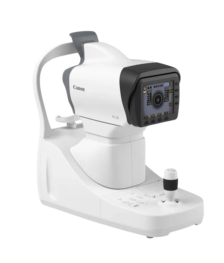
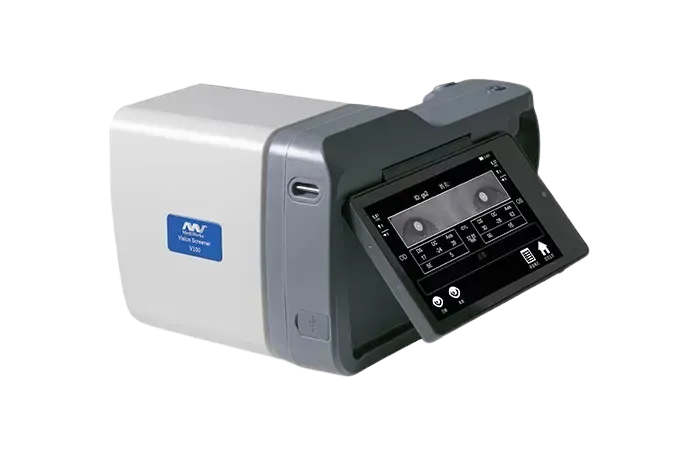
A child may not complain, because they have no experience of normal vision to compare with. A handheld screener measures both eyes from a metre away in seconds: a difference in prescription between the eyes, signs of squint and the risk of a lazy eye are picked up this way. It can be used from six months of age.
Not all of these tests are done at every examination. Your symptoms, your age and the first findings decide which measurements are added.
The devices used at an examination
How Much Does an Eye Examination Cost?
The price of eye examination varies depending on the tests to be performed. The treatment plan to be created by our specialist ophthalmologist after a detailed examination may vary from person to person.
What Happens During an Eye Examination?
The examination starts with a measurement of visual acuity. The refractive error is then measured with an autorefractometer, the pressure inside the eye is checked, and the eyelids, cornea, anterior chamber and lens are examined with a slit lamp.
Where it is needed, drops are used to widen the pupil so that the back of the eye, meaning the retina and the optic nerve, can be assessed. Depending on your symptoms, further tests such as corneal mapping, a visual field test or an OCT scan may be added.
How Long Does It Take?
A standard examination takes about 15-30 minutes. If the pupil is to be widened, it takes longer, because time has to be allowed for the drops to work.
Once the pupil has been widened, near vision is blurred and the eye is sensitive to light for a few hours. During that time you should not drive, it helps to have sunglasses with you, and coming with a companion is advisable where possible.
How Often Should You Have Your Eyes Examined?
For adults without symptoms, an examination every 2 years is advised up to the age of 40, and once a year after that. After the age of forty, for people with a family history of glaucoma and for people with diabetes, more frequent check-ups are advised, because in these cases the condition can progress without giving any warning.
In children, early examination matters because vision is still developing. Amblyopia and refractive errors may go unnoticed without the child complaining, which is why an eye examination before school age is recommended.
Before You Come In
Bring the glasses you use, any earlier prescriptions you have and a list of the medicines you take regularly. Tell your doctor if you have had eye surgery before or if you are using eye drops.
If you wear contact lenses, you may need to leave them out for a set period before the examination so that the measurements are accurate. That period depends on the type of lens, so ask when you book your appointment.