




İçeriğe geç
Kudret Eye Istanbul

Our Technology

Two things decide the outcome in eye surgery: the surgeon's experience and the equipment at hand. Below you will find the systems we use in our operating theatres and examination rooms, the manufacturers we work with, and what each device is for.

31devices under one roof

More than a typical eye clinic

Every device on this page is installed in our hospital and in daily use. We do not settle for a single instrument: we keep complementary systems so the same measurement can be confirmed by more than one method. A treatment decision then rests on measurements that agree with each other, not on a number from one device.

What this equipment means for you

  • High precision in measurements
  • A safe procedure with current technology
  • Treatment planned around your own eye
  • A short and comfortable procedure

Initial measurement devices

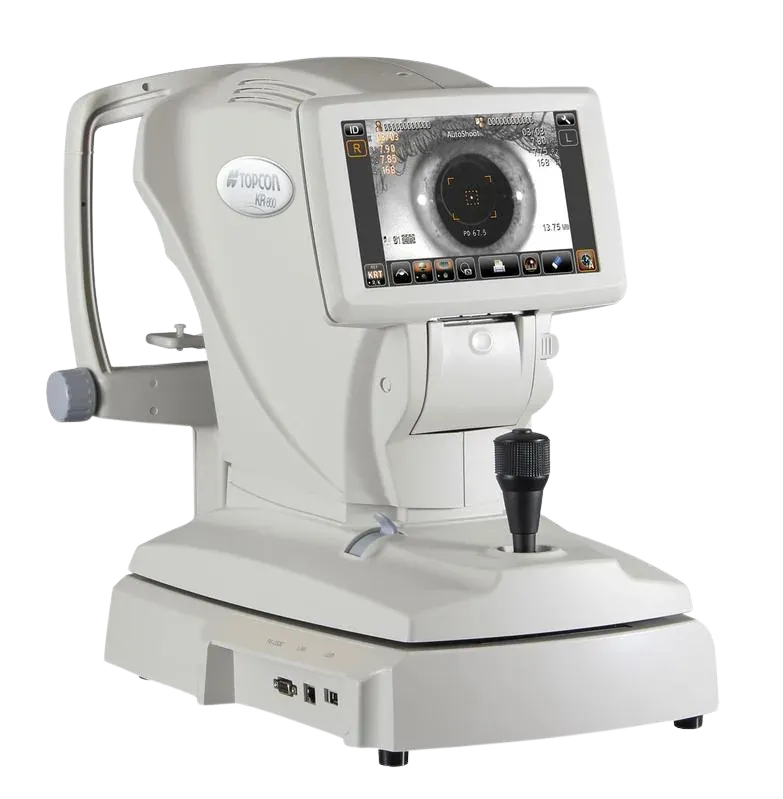
Topcon

KR-800

The device that automatically measures the eye's refractive power and corneal curvature. The first reading of myopia, hyperopia and astigmatism is taken here, and the prescription refined during the examination builds on it.

Why we chose this device

This measurement is where the examination starts. Its rotary prism method stays consistent on irregular corneas and small pupils, so the doctor fine-tunes from a reliable starting point.
Manufacturer's page

KR-800

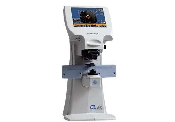
Topcon

CL-300

A computerised lensmeter that reads the prescription of the glasses a patient already wears. It measures the power, the astigmatic axis and UV protection, and recognises multifocal lenses as well.

Why we chose this device

Patients rarely know what their old prescription was. Measuring the lens itself shows how much the eye has changed over time, and the new prescription is written on top of that difference.

Manufacturer's page

CL-300

Canon

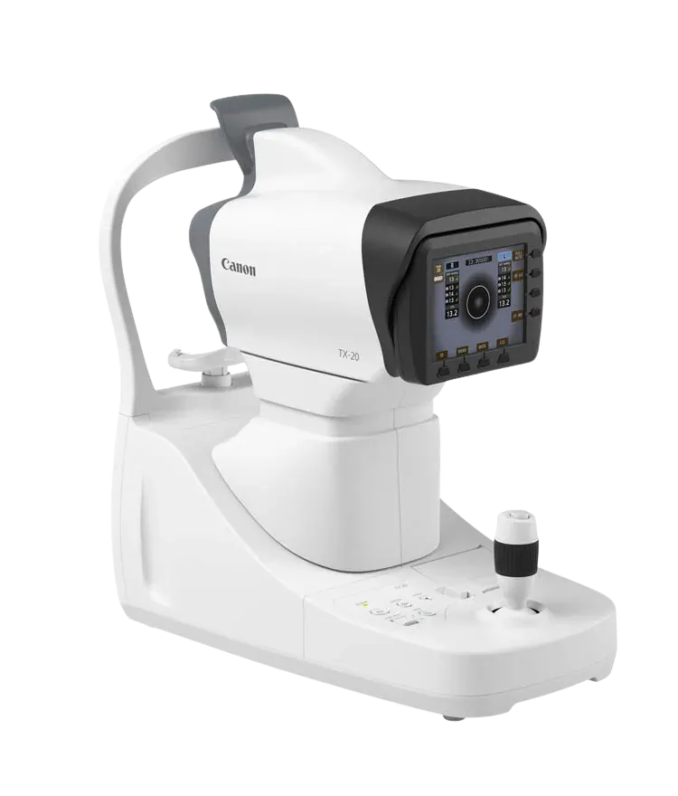
TX-20

A tonometer that measures eye pressure without contact. Nothing touches the eye but a brief puff of air, no drops are needed, and the device warns when the pressure is high.

Why we chose this device

Eye pressure is the one modifiable factor in glaucoma, so it has to be measured at every visit. Contactless measurement needs no anaesthetic drops, so the examination starts without them and the reading takes seconds.

Manufacturer's page

TX-20

MediWorks

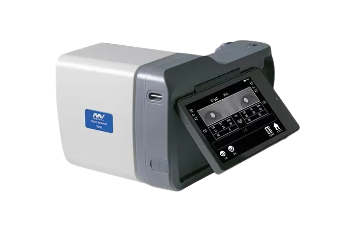
Vision Screener V100

A handheld vision screener. From one metre away it measures both eyes at once in seconds, screening for myopia, hyperopia, astigmatism, a difference in power between the eyes and signs of squint. It can be used from six months of age to adulthood.

Why we chose this device

A small child cannot sit still at a conventional instrument, yet a lazy eye is treatable when caught early and permanent when it is not. This device makes the measurement possible on a parent's lap, almost as a game.

Manufacturer's page

Vision Screener V100

Examination and imaging devices

OCULUS

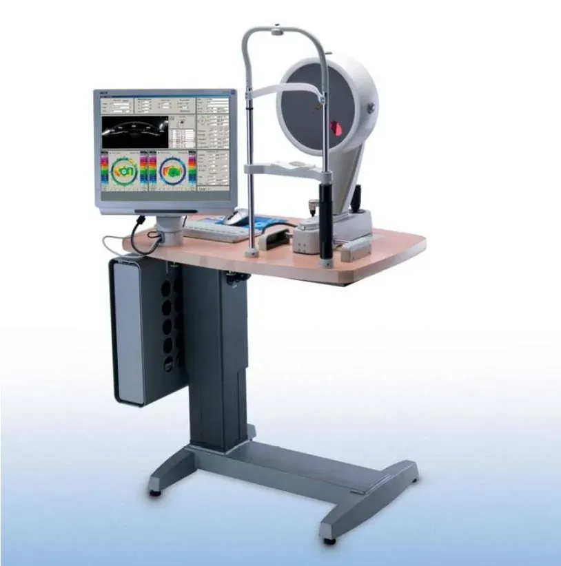
Pentacam

Scans the front and back surface of the cornea with a rotating Scheimpflug camera and builds a three-dimensional map of it. Corneal thickness, curvature and anterior chamber measurements all come from a single scan; suitability for laser treatment, lens calculation and keratoconus follow-up rest on this map.

Why we chose this device

This is the measurement that identifies an eye unsuited to laser treatment. Devices that look only at the front surface can miss early keratoconus; because the Pentacam also measures the back surface and the thickness profile, the risk shows up before anything is done to the eye. Being able to tell a patient no matters as much as saying yes.

Manufacturer's page

Pentacam

Topcon

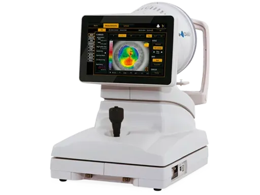
CA-800

A topographer that maps the front surface of the cornea. Alongside the curvature map it also measures the tear film, the meibomian glands in the eyelid and how the pupil changes in light.

Why we chose this device

Before laser treatment, dry eye and pupil size matter as much as the shape of the cornea. This device measures all three in one sitting, so the suitability assessment is not made on incomplete data.

Manufacturer's page

CA-800

Johnson & Johnson Vision

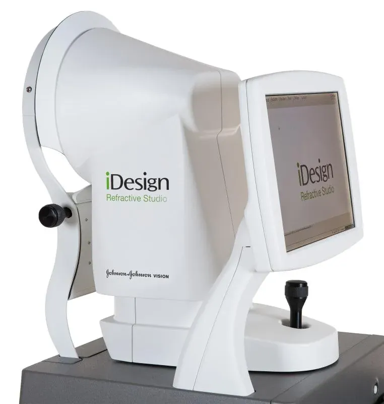
iDesign Advanced WaveScan Studio

The measurement device that captures the eye’s optical “fingerprint”. In a single scan it records refractive error at over 1,200 points, corneal topography and pupil metrics. The map is passed to the excimer laser, so treatment follows your eye’s own map rather than a standard prescription.

Why we chose this device

A spectacle prescription is a coarse summary of the eye's refractive error; this device takes thousands of separate readings across the same eye and maps them. Laser treatment is then planned around that eye's own pattern rather than an average figure, which is where night vision is won or lost.

Manufacturer's page

iDesign Advanced WaveScan Studio

Tracey Technologies

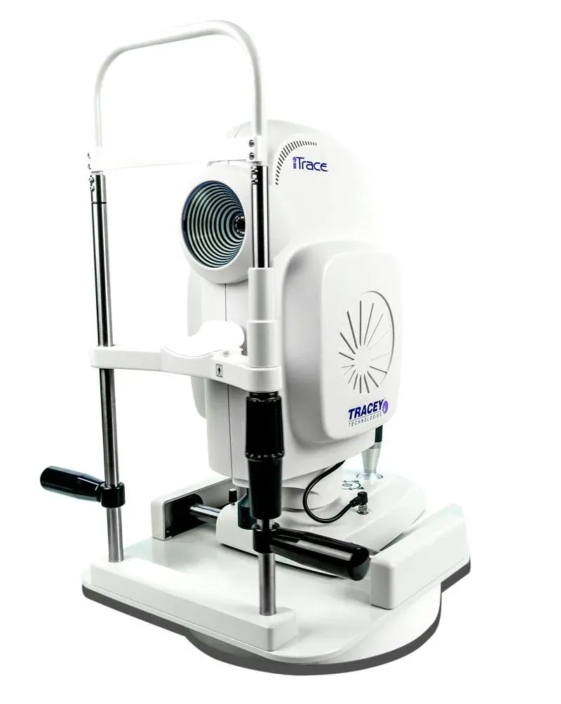
iTrace

A vision-quality analyser that splits the eye’s total refractive error into the parts coming from the cornea and from the natural lens. One of the measurements the surgeon relies on when deciding between laser and lens replacement, and when choosing a trifocal lens.

Why we chose this device

One of the methods that can tell whether a visual complaint comes from the cornea or from the lens inside the eye. That distinction changes the treatment: a corneal cause calls for laser, a lens cause for lens exchange. By ray-tracing the two separately, it keeps patients from being steered to the wrong procedure.

Manufacturer's page

iTrace

Topcon

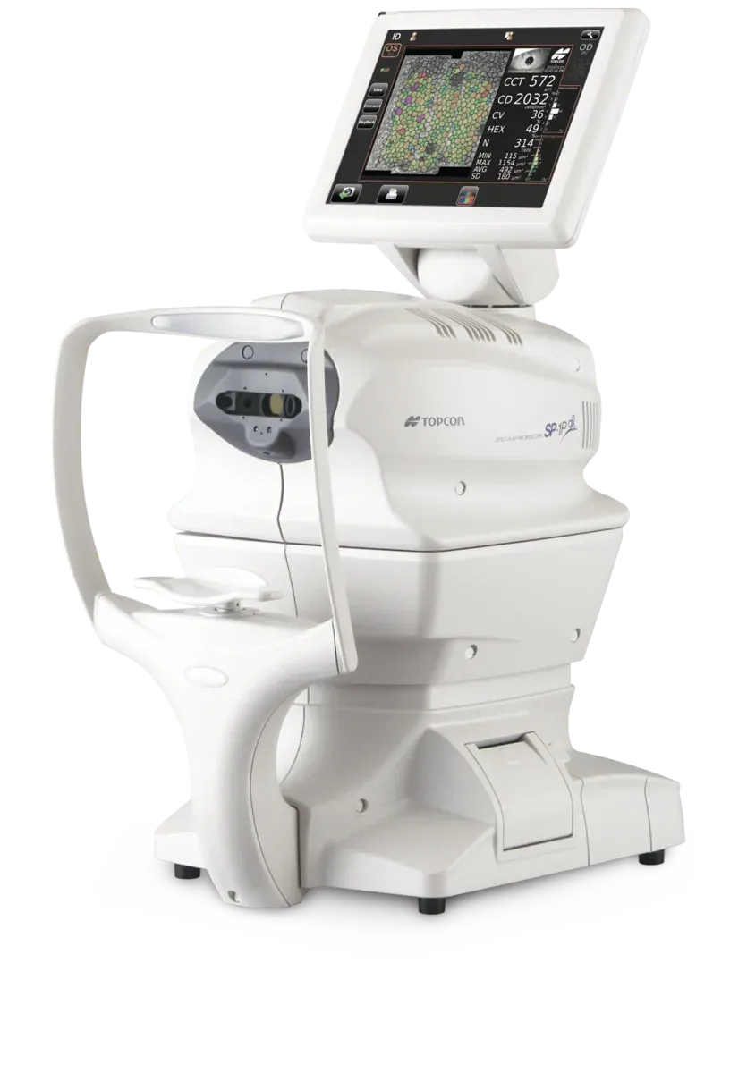
SP-1P

A specular microscope that counts the endothelial cells in the innermost layer of the cornea. These cells keep the cornea clear and do not regenerate; their number tells in advance how much strain surgery will place on the cornea.

Why we chose this device

In an eye with a low endothelial count, cataract surgery can leave the cornea hazy. Taking this measurement lets us adapt the surgical technique and energy settings to the individual patient.

Manufacturer's page

SP-1P

Topcon

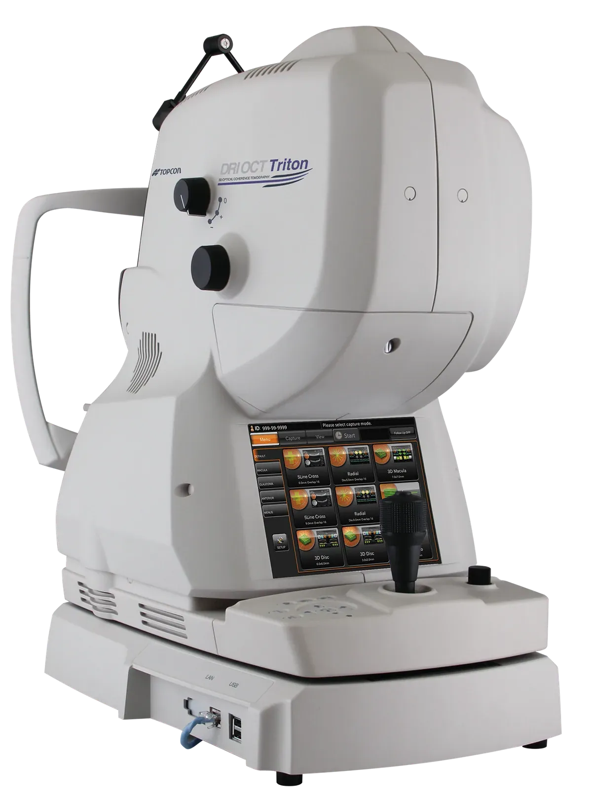
DRI OCT Triton

A swept-source OCT that shows the layers of the retina as micron-level sections. Its 1,050 nm wavelength reaches the deepest layers of the eye, down to the choroid, and the same capture also yields a colour fundus photograph. Macular disease, glaucoma and diabetic changes are followed on these sections.

Why we chose this device

The longer wavelength still reaches the retina when a cataract or bleeding inside the eye clouds the view. As the fundus photograph comes from the same capture, the patient does not sit at two separate machines.

Manufacturer's page

DRI OCT Triton

Heidelberg Engineering

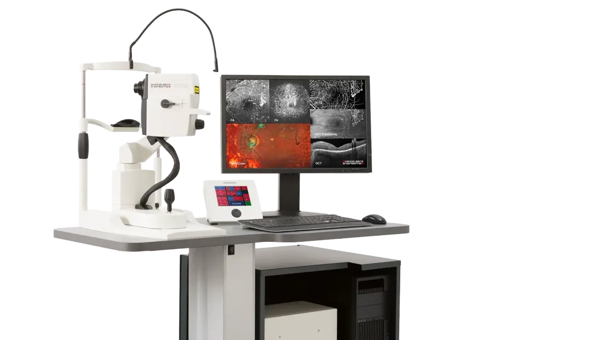
SPECTRALIS HRA+OCT

An imaging platform that combines a confocal scanning laser ophthalmoscope with OCT in one device. It captures both the surface view of the retina and its layered sections at once; angiography and autofluorescence imaging are done on the same machine.

Why we chose this device

In conditions that need follow-up, what matters is whether today's scan can be compared with the last one. This platform relocates exactly the same spot at every visit, so a difference reflects a change in the disease rather than a shift in where it was measured.

Manufacturer's page

SPECTRALIS HRA+OCT

Canon

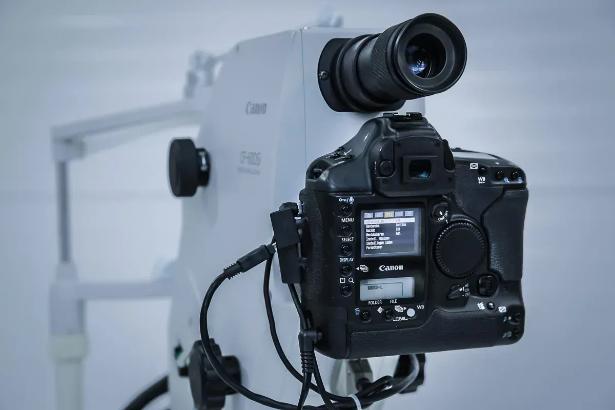
CF-60DSi

A fundus camera that photographs the retina, the macula and the optic nerve in colour. Its wide 60-degree field brings a large part of the retina into a single frame; fluorescein angiography is also performed on this device.

Why we chose this device

A colour photograph shows what cross-sections do not: bleeding, pigment change, a blocked vessel. Placing photographs taken over the years side by side is the clearest way to see which way a disease is heading.

Manufacturer's page

CF-60DSi

ZEISS

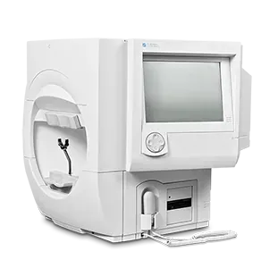
Humphrey Field Analyzer

The instrument that measures the visual field: the patient looks straight ahead and marks lights appearing around them, and the device maps where vision has weakened. It is the cornerstone of diagnosing and following glaucoma; loss can show up here years before the patient notices it.

Why we chose this device

In glaucoma the decision to treat rests on a curve spanning years, not a single test. Humphrey is the most widely used system and the one the literature is built on, so results can also be compared with older tests taken elsewhere.

Manufacturer's page

Humphrey Field Analyzer

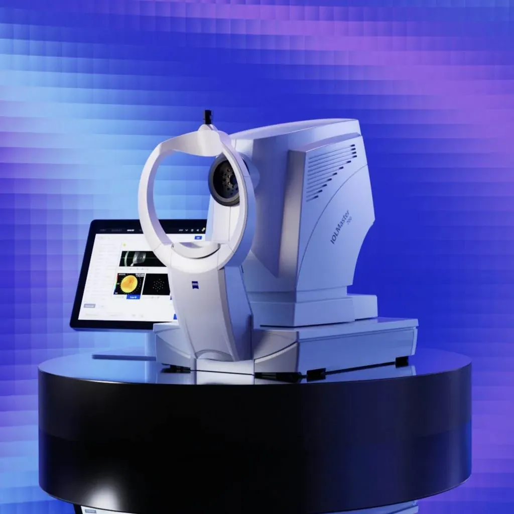
ZEISS IOLMaster 700

The optical biometer that takes every measurement of the eye before lens surgery: axial length, corneal curvature, anterior chamber depth and lens thickness. Because it produces a full-length OCT section of the eye, you can see on screen that the measurement was taken at the right point.

Why we chose this device

The result of lens surgery depends on the accuracy of the measurement taken beforehand. Because this device also shows the section it measured, a faulty reading does not pass unnoticed, and it still measures through dense cataracts.

Manufacturer's page

ZEISS IOLMaster 700

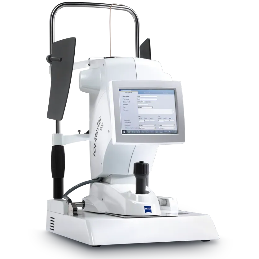
ZEISS IOLMaster 500

The device long regarded as the reference in optical biometry, used for intraocular lens calculation. It measures the axis of the eye without contact; nothing touches the patient's eye.

Why we chose this device

We keep a second biometer for redundancy: a critical measurement can be confirmed on another device, and if the two disagree the patient does not go into surgery on an uncertain number.

Manufacturer's page

ZEISS IOLMaster 500

Laser systems

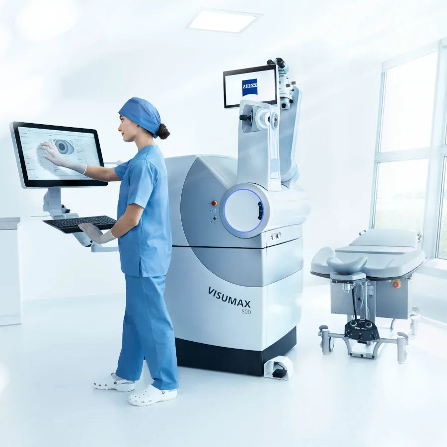
ZEISS VisuMax 800

The femtosecond laser behind ReLEx SMILE PRO. It creates a thin lenticule inside the cornea and removes it through a 2-4 mm incision; no flap is cut. Lenticule cutting takes under 10 seconds per eye and the device tracks eye movement to centre itself.

Why we chose this device

On this laser the lenticule cut in SMILE takes seconds; the shorter the eye has to be held still, the smaller the margin for error from patient movement. Its eye tracking keeps the cut on the planned axis even if the eye shifts.

Manufacturer's page

ZEISS VisuMax 800

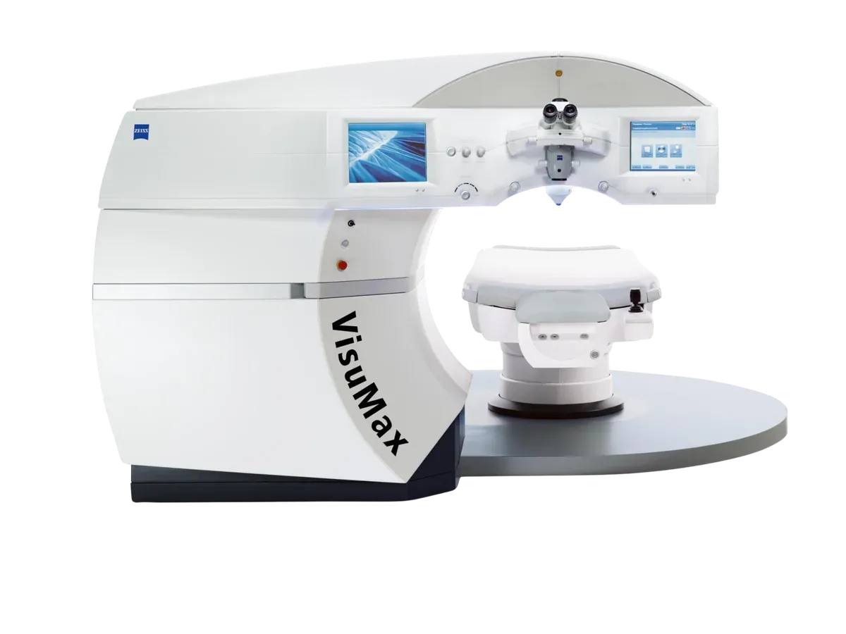
ZEISS VISUMAX 500

The femtosecond laser the ReLEx SMILE technique was developed on. It shapes a thin lens (lenticule) inside the cornea and removes it through a small incision, without raising a flap on the surface. A suction ring holds the eye with gentle contact, and the curved interface, matched to the eye's own curvature, spreads the pressure.

Why we chose this device

Most of the SMILE literature rests on results gathered on this platform, so the long-term data for the technique comes from this very device. Having it alongside the VISUMAX 800 means two femtosecond lasers: we can run two rooms on the same day, and a service visit does not push the surgery schedule.

Manufacturer's page

ZEISS VISUMAX 500

Johnson & Johnson Vision

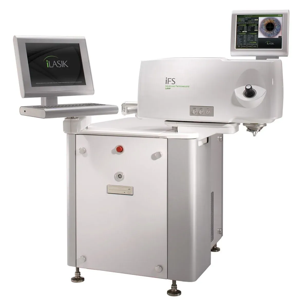
IntraLase iFS 150 kHz

The first step of iLASIK: the corneal flap is created 100 % blade-free with a femtosecond laser instead of a blade. At 150 kHz the flap forms in seconds, at the exact thickness and shape the surgeon sets, far more predictable than the older microkeratome. The device is the first link in the iLASIK platform developed by Abbott Medical Optics, today part of Johnson & Johnson Vision.

Why we chose this device

Creating the flap with a laser rather than a blade keeps its thickness uniform to the micron across the whole cornea. The 150 kHz speed shortens the step, and the bevelled edge lets the flap seat back more securely.

Manufacturer's page

IntraLase iFS 150 kHz

Ziemer

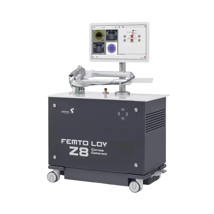
FEMTO LDV Z8

A mobile femtosecond laser usable in both corneal and cataract surgery. Its pulse energy is very low and its repetition rate very high, so cuts are formed from small overlapping spots. It can raise a flap, and in cataract surgery it opens the front capsule of the lens and fragments the lens itself. Being on wheels, the laser comes to the table rather than the patient being moved.

Why we chose this device

Opening the front capsule by hand is one of the steps in cataract surgery that most depends on the surgeon's experience; a laser-made capsulotomy comes out the same diameter and centred in every patient, which helps the intraocular lens sit where it should. That the device can be brought to the patient matters too: nobody is moved from one table to another mid-procedure.

Manufacturer's page

FEMTO LDV Z8

Johnson & Johnson Vision

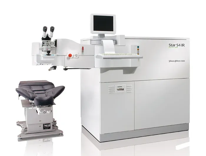
VISX STAR S4 IR Excimer

The excimer laser of iLASIK’s second step. It reshapes corneal tissue with micron precision; infrared eye tracking keeps every pulse on target even if the eye moves. Paired with iDesign measurements it delivers a fully personalised (wavefront-guided) treatment. This laser is part of the iLASIK platform developed by Abbott Medical Optics, today part of Johnson & Johnson Vision.

Why we chose this device

Infrared eye tracking measures the small involuntary movements of the eye hundreds of times a second and keeps the laser on the same spot. Its variable spot size removes only as much tissue as needed, and the less cornea spent now, the more margin remains for a later touch-up.

Manufacturer's page

VISX STAR S4 IR Excimer

Surgical equipment

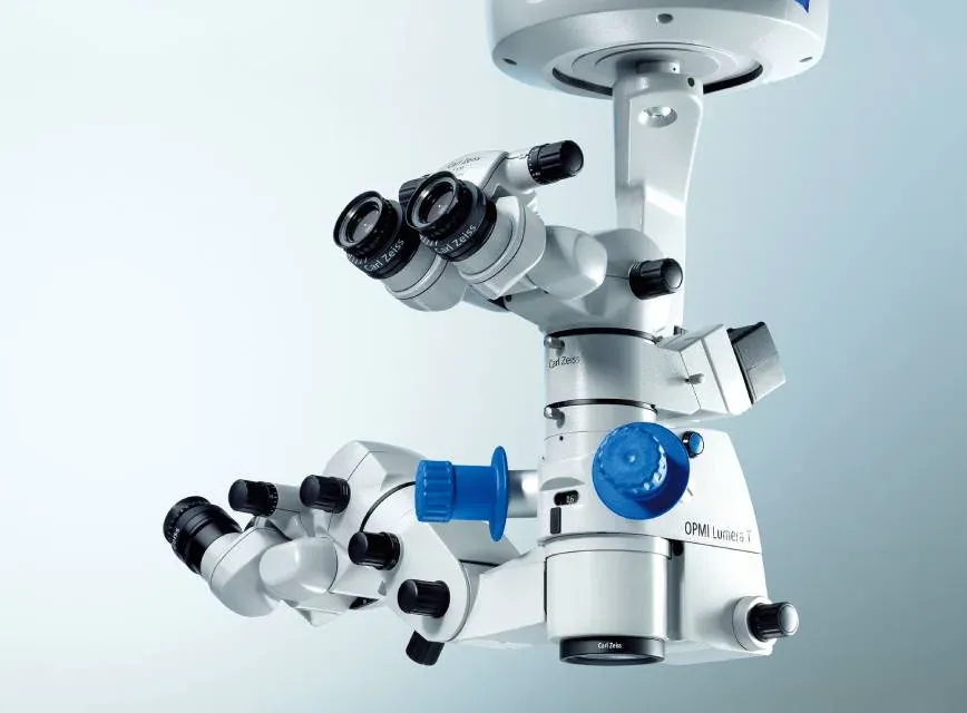
ZEISS OPMI Lumera T

The surgical microscope used in cataract and intraocular lens surgery. Its red-reflex illumination makes the edges of the lens and the capsule stand out, so the surgeon works through a high-resolution image with real depth.

Why we chose this device

Opening the capsule, the most critical step of cataract surgery, is only visible under an even red reflex. The coaxial illumination of this microscope keeps that reflex uniform across the whole eye, so the surgeon sees the edge of the capsule right to the end of the operation, even in dense cataracts.

Manufacturer's page

ZEISS OPMI Lumera T

Alcon

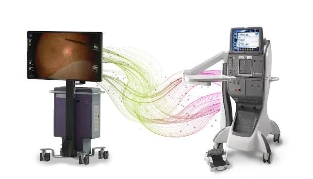
CONSTELLATION Vision System

The surgical system used in retinal and vitreoretinal operations. It measures the pressure inside the eye throughout surgery and balances it automatically, and it controls the cutter speed and the suction independently of each other. Laser ports are built in.

Why we chose this device

The main risk in retinal surgery is the pressure inside the eye swinging during the operation. A system that measures and balances it continuously keeps the retina stable throughout.

Manufacturer's page

CONSTELLATION Vision System

Alcon

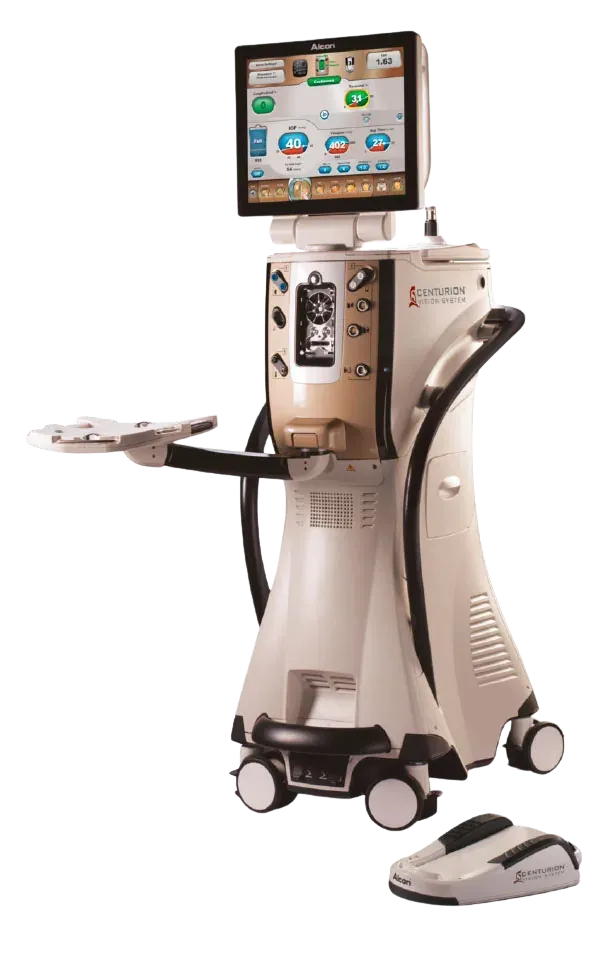
Centurion Vision System

Alcon's Centurion Vision System is the surgical platform that breaks up and removes the natural lens with ultrasound (phaco) in cataract and multifocal lens surgery. Its active fluid management keeps eye pressure stable throughout the procedure, so surgery is shorter and gentler on the tissue.

Why we chose this device

In cataract surgery a sudden drop in intraocular pressure means the anterior chamber collapsing, and preventing that moment is decisive for safety. This system senses the pressure at the tip of the handpiece and balances it there, so it can absorb the surge the instant an occlusion clears.

Manufacturer's page

Centurion Vision System

Johnson & Johnson Vision

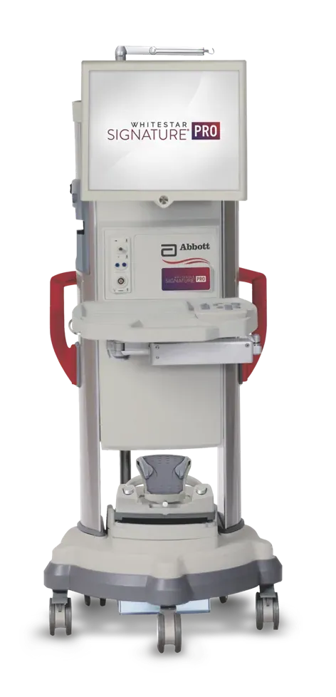
WHITESTAR SIGNATURE PRO

The phacoemulsification system used in cataract surgery: it breaks up the hardened lens with ultrasound energy and removes it from the eye. It senses occlusion by itself to protect the pressure inside the eye and can switch between peristaltic and venturi pumps during the operation.

Why we chose this device

The less ultrasound energy used, the less the cornea is affected and the sooner vision clears. This system delivers energy in pulses, which means less total energy for the same cataract.

Manufacturer's page

WHITESTAR SIGNATURE PRO

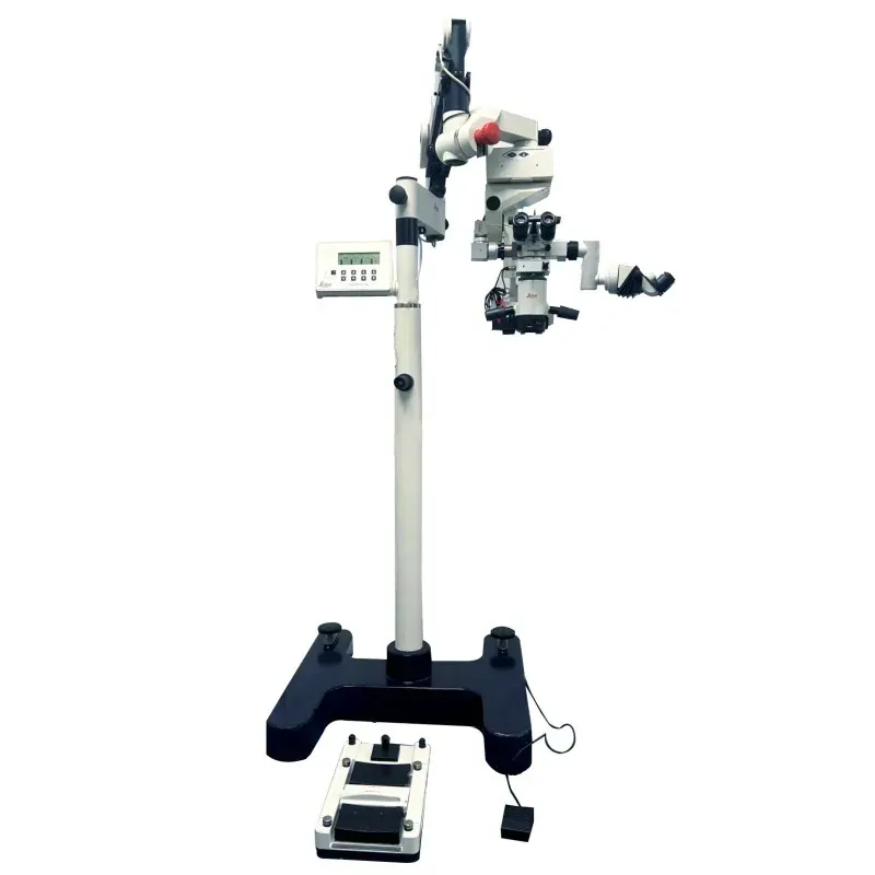
Leica M841

The surgical microscope used in eye operations. Its APO OptiChrome optics render colours faithfully and the OttoFlex illumination keeps the red reflex even throughout surgery, so the surgeon and the assistant see the same image at the same clarity.

Why we chose this device

In cataract surgery the edge of the capsule is only visible under an even red reflex; when illumination drops, the surgeon has to judge the most delicate step by guesswork. Keeping a second theatre microscope also lets us operate in two rooms at once.

Manufacturer's page

Leica M841

Intraocular lenses

Johnson & Johnson Vision

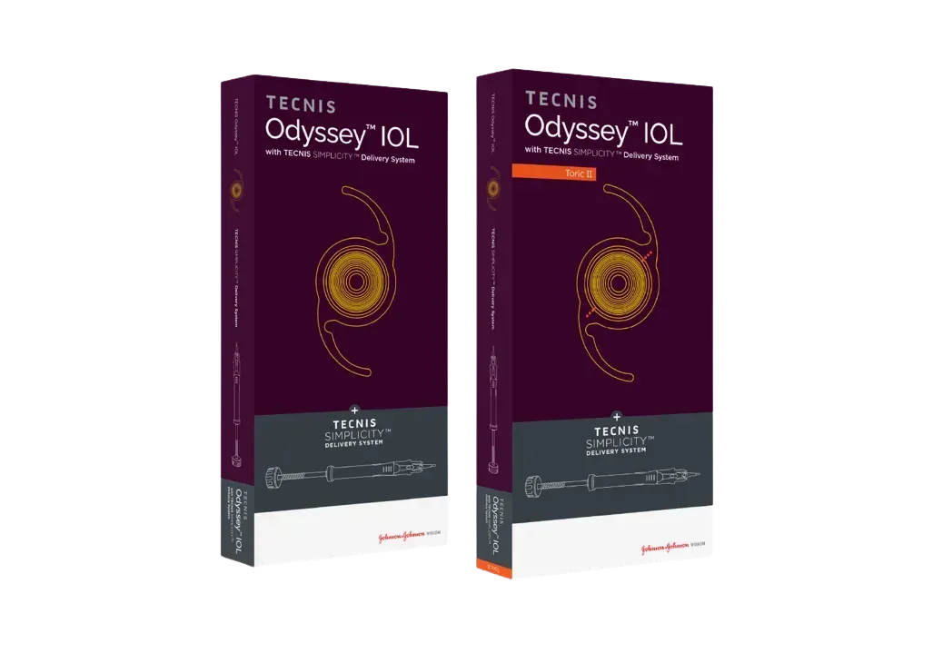
TECNIS Odyssey

A multifocal intraocular lens that replaces the natural lens in cataract and refractive lens surgery. It is designed to remove the need for glasses at all three distances, near, intermediate and far, delivering them as one continuous range with no gap at the range of a computer screen or a dashboard.

Why we chose this lens

The complaints heard most often about multifocal lenses are haloes at night and a gap between the focal distances. We put Odyssey first because of the progress it makes on both: it is designed to scatter less light, which helps night driving, and its continuous range aims at glasses-free vision at all three distances.

Manufacturer's page

TECNIS Odyssey

Alcon

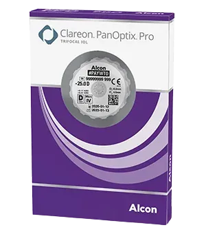
Clareon PanOptix

A trifocal intraocular lens: it divides the incoming light between three distances, near, intermediate and far, and is designed to remove the need for glasses at all three. The Clareon material was developed to reduce the glistenings and surface haze that can appear on a lens over the years.

Why we chose this lens

One of the trifocal families with the longest follow-up data, so it is well understood which patients do well with it. Its move to the Clareon material matters for the twenty or thirty years the lens stays in the eye: an intraocular lens is implanted once, and exchanging it later is not straightforward.

Manufacturer's page

Clareon PanOptix

Johnson & Johnson Vision

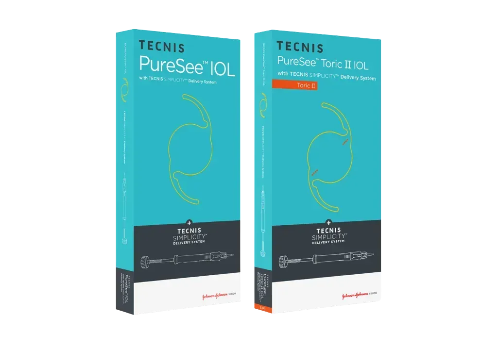
TECNIS PureSee

An extended depth of focus (EDOF) intraocular lens. Rather than splitting light into separate focal points, it stretches the zone of focus, giving distance and intermediate vision as one continuous range. Its surface is smooth, without diffractive rings.

Why we chose this lens

A better fit than a trifocal for patients who drive a lot at night or tolerate haloes poorly. Without diffractive rings, night image quality stays close to that of a monofocal lens; in exchange, reading glasses may still be needed up close. We discuss that trade-off with the patient beforehand.

Manufacturer's page

TECNIS PureSee

Alcon

Clareon Vivity

An extended depth of focus (EDOF) intraocular lens. A very small step in the centre of the lens reshapes the wavefront of the incoming light and stretches the zone of focus, without the diffractive rings that split light into separate focal points.

Why we chose this lens

Trifocal lenses are usually avoided in eyes with an added problem such as macular disease or glaucoma, because splitting the light lowers a contrast that is already reduced. Vivity does not split the light, which makes it one of the options for these patients; we decide by looking at the retina and optic nerve measurements.

Manufacturer's page

Clareon Vivity

Rayner

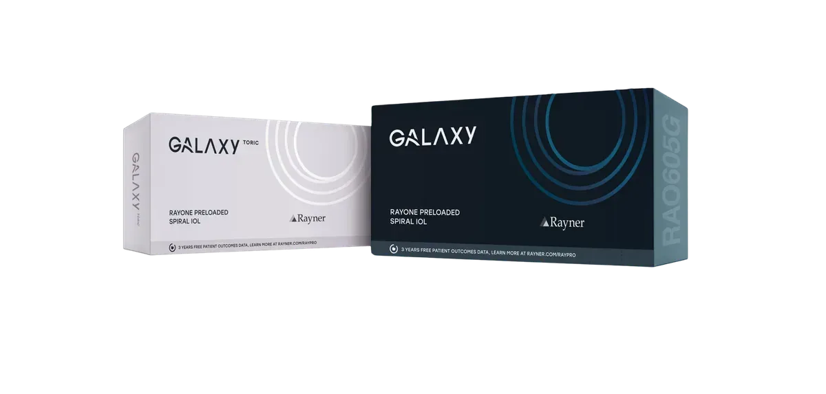
RayOne Galaxy

A trifocal intraocular lens whose surface carries a continuous spiral instead of stepped rings, designed to remove the need for glasses at all three distances, near, intermediate and far. The spiral is designed to direct light without passing it over steps. The lens arrives preloaded, already folded inside its own injector.

Why we chose this lens

Stepped rings are the main source of night-time haloes in multifocal lenses. The spiral design removes those steps and aims to reduce the halo effect. The lens also arrives preloaded, which removes the step of loading it by hand in theatre and the risk that comes with it.

Manufacturer's page

RayOne Galaxy

STAAR Surgical

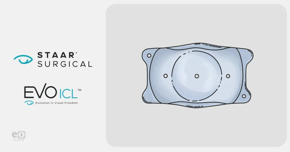
EVO ICL

A lens placed inside the eye, behind the iris, while the natural lens stays where it is. It is made of Collamer, a material compatible with the eye's own tissue. No corneal tissue is removed, and if needed the lens can be taken out and the eye returns to its previous state.

Why we chose this lens

Laser is not an option for a patient with a thin cornea or a prescription above the laser limit; for them this lens is the main route to life without glasses. That it is reversible matters too: laser removes corneal tissue that does not grow back, whereas this lens can be taken out.

Manufacturer's page

EVO ICL

We keep renewing our equipment

Devices in eye care change quickly, and a newer system often means a shorter, more comfortable or more precise examination. We have renewed our equipment regularly since the day we opened, bringing new-generation systems into the clinic. This page is not fixed either: when a new device arrives, it is added here.

FREE CONSULTATION

Your appointment request has been received successfully.

We will be in touch with you as soon as possible.

If your details are not correct, please send a new request.

Your cookie choice

Cookies needed for the site to work are always on. With your permission we also use Google and Meta cookies to measure visits and show you relevant ads. You can change your choice at any time with the "Cookie settings" link at the bottom of the page.Detailed Information

Suitability test

Which treatment suits me?

A few short questions to show you which treatments would be discussed at your examination. At the end we will call you for a free consultation.

Book a Free Consultation

FREE CONSULTATION

Site search

Can't find what you're looking for?

We are just a phone call away.