




Eye Diseases
What is Retinal Detachment?
- Kudret Eye Istanbul
- Last updated:
- 6 min read
- Medically reviewed by Op. Dr. Mehmet Ali Özdil
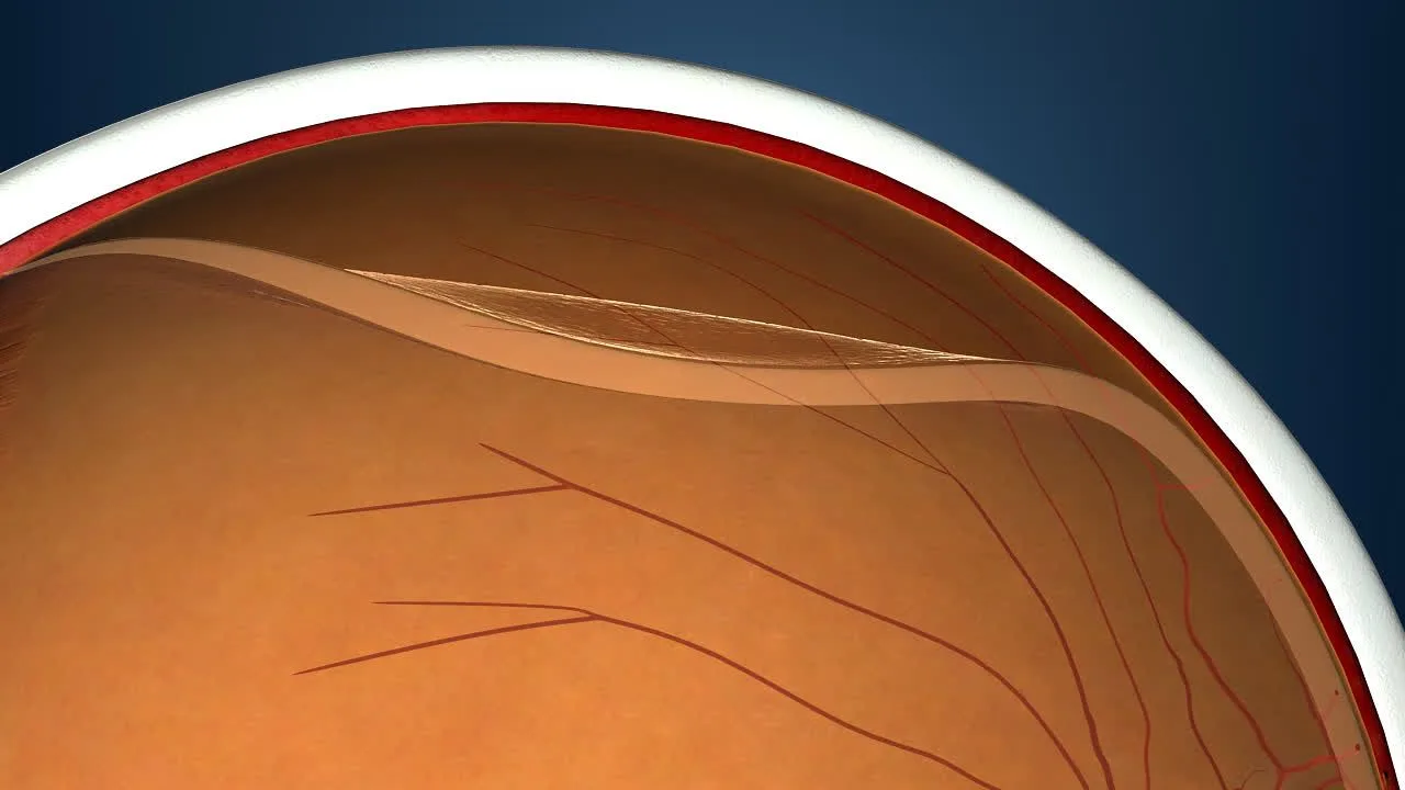
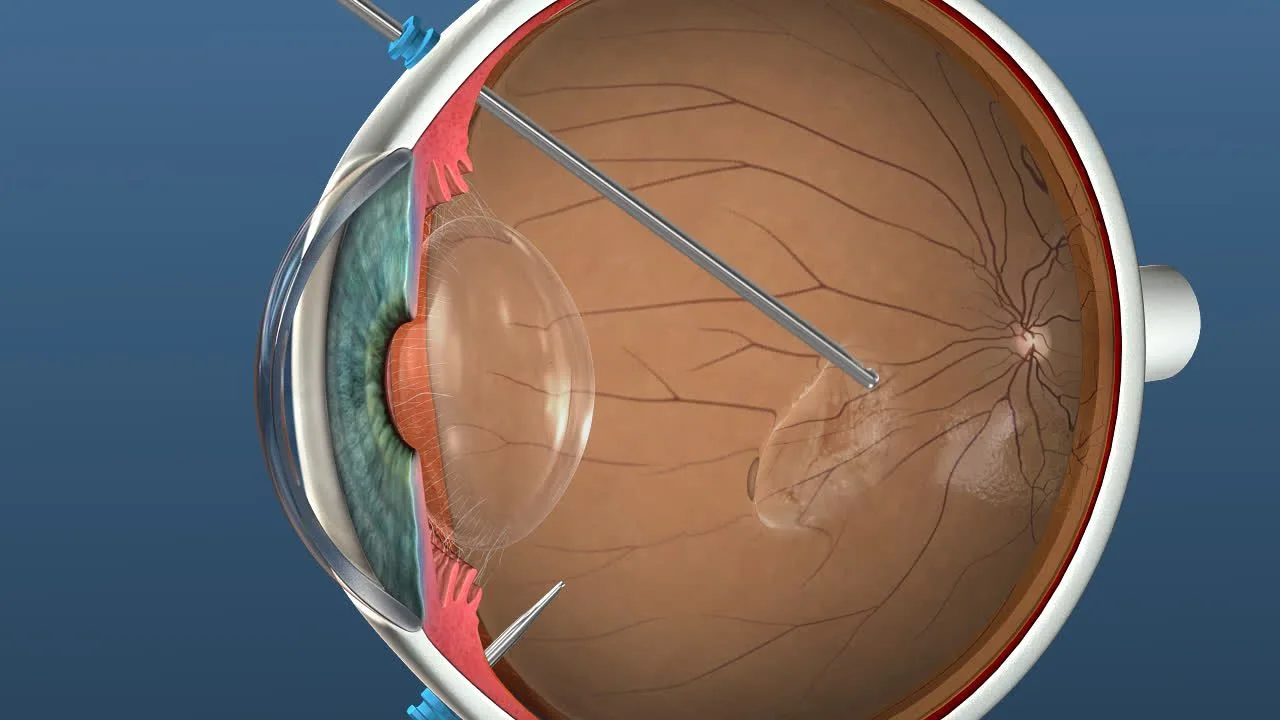
The question of what retinal detachment is falls among those asked most by people facing this serious condition. Retinal detachment occurs as fluid gathers beneath the light-sensitive retinal layer and the sensory retina separates from the pigment epithelium. It is a very serious condition. The type seen most often is rhegmatogenous detachment, which generally forms as vitreous fluid leaks through a retinal tear that has not been sealed with a laser. It appears in situations such as advanced age, high myopia (minus six dioptres and above) and injuries to the eye.
Retinal detachment shows itself in many ways. Vision declines and flashes of light occur in the eye. The warning signs are flashes of light (photopsia), a sudden increase in black spots or floaters, the sense of a curtain across the field of vision, seeing straight lines as broken, or loss of central vision. There is generally no pain; burning in the eye is not typical of detachment. If it is left untreated it leads to sight loss over time. This condition can be diagnosed in many ways. What matters is seeing a specialist in time, and early diagnosis always matters a great deal.
Retinal detachment is a condition occurring in the retina. The retina allows the eye to perceive light and carries that light to the brain through the optic nerves. This structure is attached to the pigment epithelium. If it separates from the inner part of the eye, the condition is called retinal detachment.
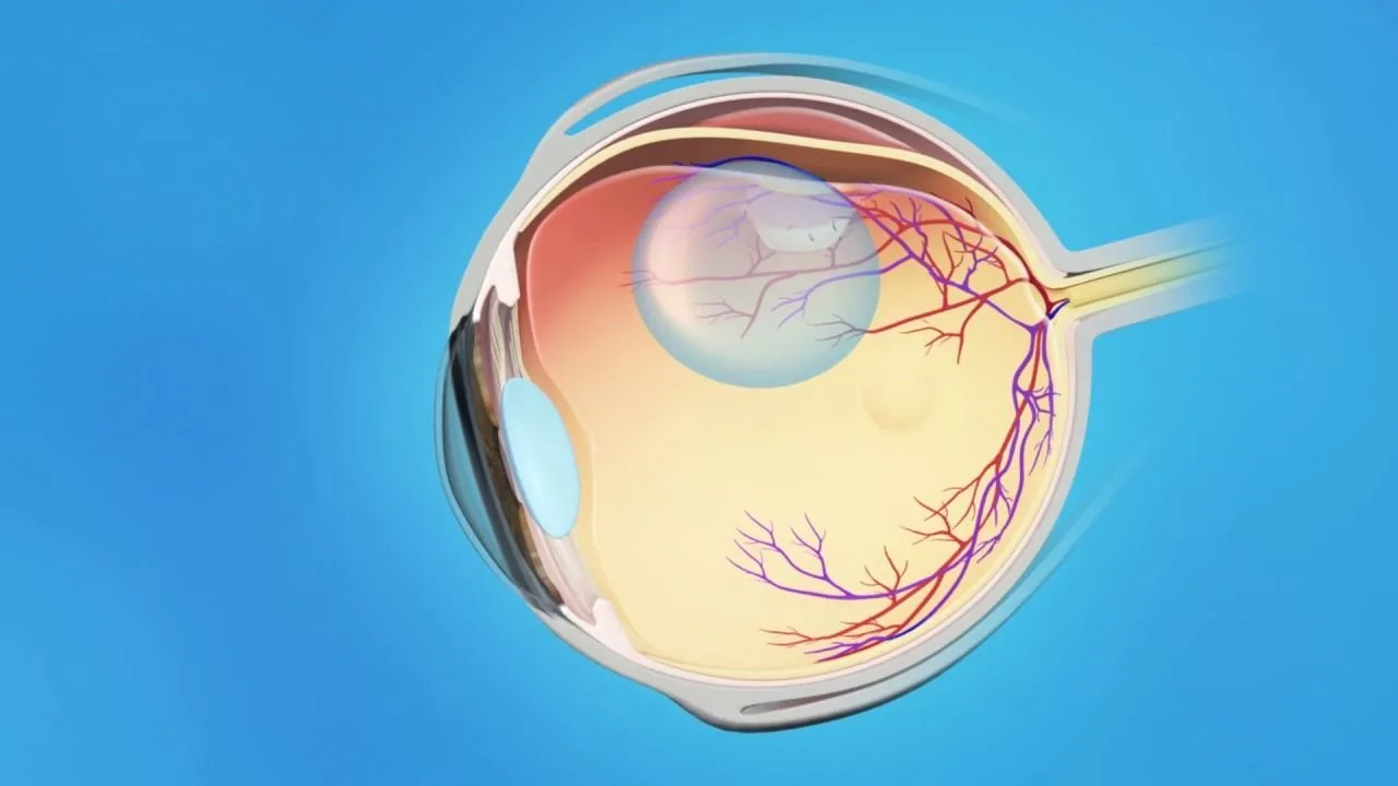
Retinal detachment is often preceded by a retinal tear, a break in the retina while it is still attached to the inner part of the eye. If a retinal tear is left untreated, the structure separates completely from the inner part of the eye over time and retinal detachment follows. If that situation is left untreated for a long time, the function of sight can no longer be carried out and permanent damage can follow. These situations show themselves in different ways. The symptoms should be taken seriously and an ophthalmologist seen as soon as possible.
This condition can occur through the deterioration that comes with advanced age, through damage to the eye, through certain eye operations and through high myopia. Retinal tears are usually sealed with laser treatment. Where there is retinal detachment, however, surgery is necessary. It shows itself through perceiving objects at different sizes, seeing broken lines and flash-like bursts in the eye. If one or more of these situations is present, a doctor should be consulted immediately.
What Causes Retinal Detachment?
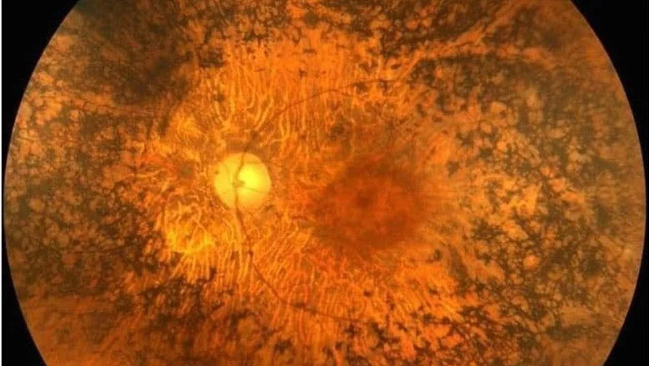
What causes retinal detachment is another important question. People with myopia of six dioptres and above, and those whose eye structure has deteriorated with age, are more predisposed to this condition. Certain problems and complications can also occur after cataract surgery, which can lead to retinal tears. The retina of people with high myopia is thinner than normal, which increases the risk of a tear considerably. Retinal problems also arise as the structure of the eye deteriorates with advanced age. In some situations the cause of the detachment cannot be identified, which is why early diagnosis matters so much. So that serious sight loss is not experienced at advanced stages, a specialist ophthalmologist should be seen if the following are observed:- High myopia of minus six dioptres and above
- A previous retinal detachment in the other eye
- Injuries to the eye
- Eye surgery
- A family history of retinal tears
- Weakening of the retina
- Diabetes
- Complications following cataract surgery
- A reduction in the vitreous fluid
- The retina becoming taut
- Structural problems present in the eyeball
- Advanced age
Treatment of Retinal Detachment
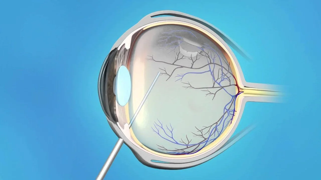
Retinal detachment is treated according to the situation. A retinal tear caught before detachment develops can be sealed with laser; once detachment has developed, the treatment is surgery. Vitrectomy is among the treatments used in retinal tears and retinal detachment. The fluid within the eye is replaced with a new fluid. In another method, where there is a tear in the retina, silicone is placed on the outer part of the eye and the retina is attached back in place. In another method, gas is injected into the eye so that the retina is returned to its former position. The doctor's advice should be followed after surgery and check-ups attended regularly. Where medication is involved, it should always be taken on time.
Retinal Surgery
There are many types of retinal surgery, determined according to the state of the retina. The tissue called the retina is a very important structure in allowing the eye to carry out the function of sight. This layer, onto which light is projected, carries that light to the brain through the optic nerves, and the function of sight is thereby carried out. Alongside holding millions of nerve cells, this part carries light to the optic nerves through those cells. The brain processes that light and those signals and turns them into an image. The retina is a delicate structure, and the structures around it are extremely delicate as well. Problems occurring around the retina can affect it directly. Where this structure tears or detachment occurs, surgery can be turned to. Retinal operations include:- Scleral buckle
- Argon laser
- Vitrectomy
- Sight loss is prevented.
- Microsurgery allows very delicate procedures.
- The retina is placed back healthily and the tissues repaired.
Recovery After Retinal Surgery
Recovery after retinal surgery matters a great deal. Although patients are generally not admitted, they can be kept under observation for a night depending on the doctor's decision. The things to observe during this period are:- Avoiding lifting heavy objects and heavy sport
- Where gas has been injected into the eye, lying face down for a period as the doctor advises, and not flying
- Paying attention to the cleanliness of the operated area
- Keeping the eye covered for several days and avoiding contact with water
- Protecting the operated eye from blows and pressure
- Taking the medication the doctor has given exactly on schedule