


 Türkçe
 English
 Deutsch
 Nederlands
 Italiano
 Español
 Français
 Български
 Română
 Русский
 العربية

Kudret Eye IstanbulEye Diseases
What is a Visual Field Test?
A visual field test measures how well peripheral vision is working. It shows how far you can see to the sides, below and above the object you are…

Treatments
Eye tests are short assessments that measure different functions of the eye, from visual acuity to colour vision and the visual field. They are used together during a comprehensive examination and reveal refractive errors as well as the first signs of eye disease.
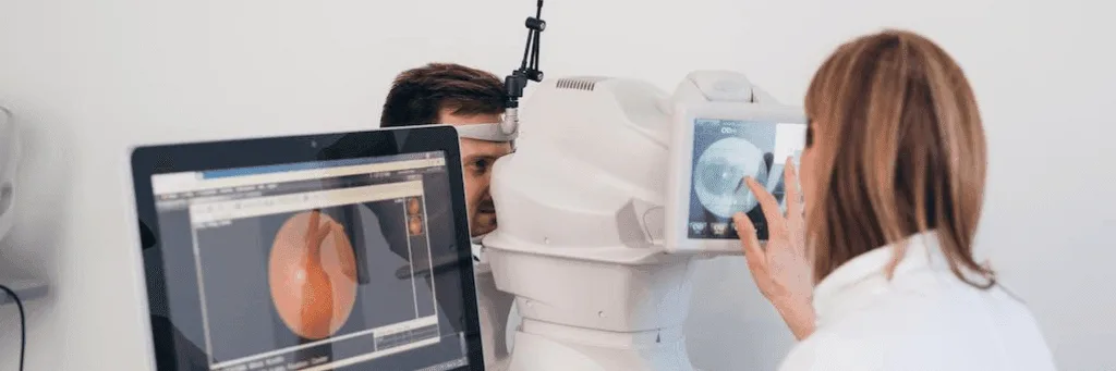
An eye test is a series of tests used to evaluate your eyes and eyesight. These tests are performed by ophthalmologists during a comprehensive eye examination. Eye tests use different methods to assess your visual acuity, colour vision, the function of your eye muscles, eye pressure and more. Common eye tests include the following.
A visual acuity test is a test that measures how clearly the eyes can see at a certain distance. This test is used in the diagnosis of refractive errors such as myopia, hyperopia, astigmatism as well as eye diseases such as macular degeneration, cataracts, glaucoma.
The visual acuity test is performed by an ophthalmologist in a clinical setting. The test proceeds as follows:
Visual acuity is expressed as a fraction of two numbers. The first number is the distance at which the test is done and the second is the distance at which a normal eye reads the same line. 6/6 vision means you read at 6 metres what a normal eye also reads at 6 metres, while 6/12 means a normal eye reads at 12 metres the line you can only read at 6. People with visual acuity impairment may be offered options such as glasses, contact lenses or surgical treatment.
This test shows how the eye is performing today, but not why. A line lost on the chart can come from an uncorrected prescription, from a cataract, from the macula or from the optic nerve, and the treatment for those four has nothing in common. Acuity is therefore where the examination begins rather than where it ends: it says how much has been lost, and the measurements that follow say where from.
Its second value shows up in follow up. If acuity is dropping while the prescription has not changed, the problem is not in the glasses but in the eye itself. In children, a difference in acuity between the two eyes is the first sign of a lazy eye, and that difference only appears when each eye is measured on its own.
Colour blindness, also commonly known as daltonism, is a visual impairment in which the eyes have difficulty perceiving certain colours or shades of colour. The most common type of colour blindness is red-green colour blindness, which causes difficulty distinguishing between the colours red and green.
Colour blindness testing is a series of diagnostic tests used to assess a person's ability to perceive colours. Specifically, these tests measure whether a person can correctly distinguish certain colours or the ability to see shades of certain colours.
The colour blindness test is carried out in a well-lit room and both eyes are tested separately. The doctor will show you a series of coloured shapes or patterns and ask you how you see them. In the treatment of colour blindness, some assistive devices, such as contact lenses and special glasses, help people with colour blindness to better distinguish colours.
Congenital colour blindness stays the same for life and does not progress. That is where the value of the test lies: if colour perception has changed in an adult who used to see colour normally, the finding is no longer about colour. Acquired loss of colour vision, particularly in the red and green range, is one of the early signs of optic nerve disease, and patients rarely notice it themselves.
The test also has an occupational use. Aviation, driving, seafaring and electrical work all require certification, because telling colours apart is directly tied to safety. In children, early diagnosis matters for schooling: a child who cannot separate colour coded material usually does not say so.
The Amsler Grid test is used to diagnose diseases affecting the centre of vision, such as macular degeneration. It is performed using a simple grid of lines. The doctor tests each eye of the person separately.
It pays attention to whether the lines on the grid are straight, whether any area appears distorted, blurred or dark in colour.
A grid of straight lines has one purpose: to make visible the distortion a patient would otherwise explain away. Lines that bend, or a patch in the middle of the grid that is blurred or missing, point straight at the macula. Patients usually do not notice the change, because with both eyes open the healthy eye fills in the missing part. Covering one eye at a time is what exposes it.
The other advantage of the test is that it can be done at home. People who have macular disease or are at risk of it can check the grid themselves at regular intervals and catch a new distortion within days. Those days matter, because in wet age related macular degeneration how early treatment starts decides the outcome.
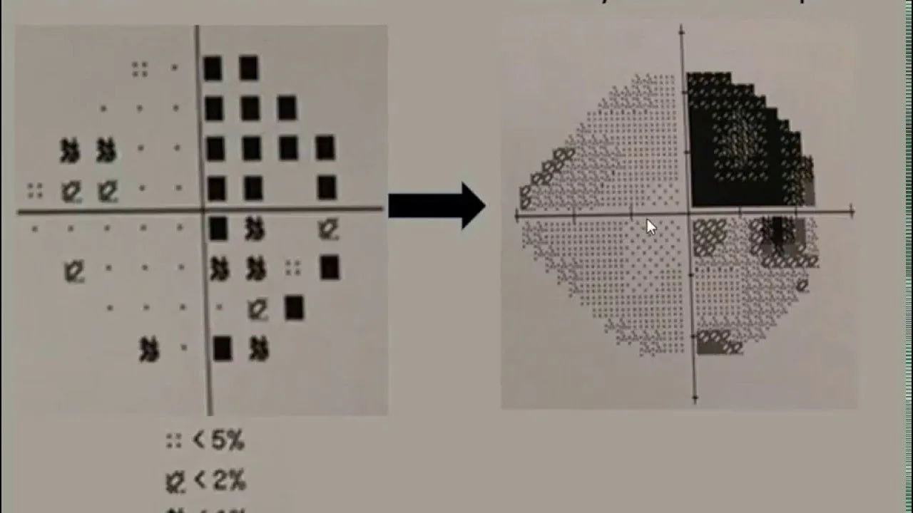
A visual field test is used to determine how much of our visual field we can see. It helps to assess how the brain processes the signals it receives from the visual nerves. It is used to diagnose diseases that can narrow the visual field, such as glaucoma, retinitis pigmentosa and brain tumours. The test is performed by an ophthalmologist using a special device.
Peripheral vision can be lost gradually while central vision stays sharp, and the brain fills in the missing area so convincingly that people go years without suspecting anything. The loss is usually noticed only in daily life: missing a car approaching from the side, catching a door frame, misjudging a step. The test maps where that loss is.
It is the cornerstone of diagnosing and following glaucoma. Eye pressure shows the risk and OCT shows the structural thinning of the optic nerve, while the visual field shows what that damage actually costs in everyday sight. Repeating the test at set intervals is also the only way to judge whether treatment has stopped the loss.
The shape of the loss says something about its cause as well. Loss on the same side in both eyes points not to the eye itself but to the visual pathways or the brain, so the test sometimes gives the first sign of a condition outside the eye. A visual field of a certain width is also required for a driving licence and for some occupations.
The Duochrome test is an eye test that ophthalmologists use to check whether a prescription is focused exactly on the retina. The test is done as follows.
By comparing the clarity of letters or symbols on red and green backgrounds, the patient's ability to see clearly in both colours is evaluated.
Red and green light focus at slightly different depths in the eye: red falls a little behind the retina and green a little in front of it. Comparing how sharp the letters look on the two backgrounds therefore says whether the prescription sits exactly on the retina. If the letters on red are sharper the correction is slightly under, and if those on green are sharper it is slightly over.
The test is not used to look for disease but to settle the final quarter of a dioptre. It works in people who are colour blind too, because the question is not which colour is seen but on which background the letters are clearer.
The tests on this page are ones you take part in: you read, you compare, you say what you see. Alongside them are measurements the devices take without asking anything of you, and they answer different questions: eye pressure, the thickness and map of the cornea, the layers of the retina, the cell count in the cornea.
The two do not replace one another. A raised eye pressure or a macula that has started to thin gives no symptom you could report, which is why these are measured rather than asked about. Not every test is done at every examination: your symptoms, your age and the first findings decide which measurements are added.
What each of the device measurements is for, and which device takes it, is set out on our general eye examination page.
Eye test price varies depending on the test to be performed. The treatment plan to be created by our specialist ophthalmologist depending on the detailed examination result may vary from person to person.
At the end of your eye test you are given a prescription, usually valid for two years. Its numbers and abbreviations describe the condition of your eyes and the correction you need. Tap a term and its column lights up in the table.
Sphere (SPH)
The main correction for short- or long-sightedness. A minus (−) means you struggle to see far away, a plus (+) means you struggle up close. The larger the number, the stronger the prescription.
Sample prescription
Over 24 years in eye care: three eye hospitals and a general hospital, a team of specialist surgeons and technology renewed every year.
Hundreds of thousands of successful operations and a name trusted across generations.
World-leading equipment for SMILE PRO, iLASIK and trifocal lenses.
Every treatment is performed by a surgeon who specialises in it.
Follow-up checks and, if ever needed, enhancement are our responsibility.
Outstanding care from start to finish. The team ensured I was fully briefed before and upon my arrival, and I felt reassured throughout the entire procedure. Jem, Dr Mehmet Ali Ozdil and Zuhal delivered exceptional service and made me feel completely at ease during my Femto‑LASIK surgery. Note, following their simple aftercare instructions is also key to recovery.

They made the process of coming to a country where I don’t speak the language and having eye surgery (frightening enough in your home town) very easy indeed. I had a wonderful interpreter with me through the entire process every day, she made everything so easy. First class facilities, first class staff and a very convenient location.
A very kind and professional medical team! The surgery was completely straightforward and was completed relatively quickly. The entire team, and especially Zuhal, my patient coordinator, was always available, incredibly helpful, and genuinely pleasant throughout the whole process. She stayed by my side from start to finish, explained everything thoroughly, and always made sure I understood what was happening. The doctor worked in a very calm and professional manner, and I never felt uncertain or uncomfortable at any point. There were three fresh meals each day: breakfast, lunch, and dinner, and they were all very enjoyable. The hospital rooms were also very clean and well maintained. Overall, I had a very positive experience with this hospital. I’m truly grateful to the entire team and would wholeheartedly recommend this hospital to anyone.
Hello, so I went to Optical Express in UK but they quoted me an insanely high price for LASIK Surgery. After conducting research on multiple different hospitals and eye surgery places in Turkey. I settled on Kudret Goz. Throughout the entire period they were helpful in answering all my questions and very supportive. They have very experienced doctors who perform LASIK surgery multiple times a day for 20 years plus using the latest Johnson and Johnson technology. As soon as I landed at the airport they picked me up, dropped me at the hospital where I had my own comfortable room. They provided breakfast, lunch and dinner. In the morning they conducted tests to check my vision and if I’m feasible for the surgery, the same evening the surgery happened. I was nervous but It lasted 5 - 10 minutes and rested in my room that evening taking eye drops and disinfect eye drops every hour. The next day I woke up and my vision had become incredibly better, with every passing hour it got better and better. Within 24 hours my vision was incredibly clear and I was amazed. Thank you for operating the surgery and truly providing a life changing experience. For the price and quality of service Kudret Goz is unrivalled.
I have just returned from having Lens transplantation at Kudret Eye Hospital Istanbul. I was a lone traveller who was terrified at the prospect of eye surgery even having done my research. I have to say ALL the staff at Kudret were absolutely amazing. From 1st contacting them the communication was excellent no question was too much and always received a very prompt response. I was met at the airport in the middle of the night and taken straight to the hospital, was shown to my room. I felt very safe. I had numerous test the following day and then a consultation with the Doctor who advised me of my best implants. There was always someone with me who was able to translate anything said. I had my 1st op that afternoon again I felt safe and very confident with the professionalism of all the staff. Surgery took approximately 20 mins. I had no pain and the next day was able to see near intermediate and distance although not 100% but without glasses. After further checks I then went in for my second surgery. Again all went smoothly. The following day sight had improved even more in my 1st eye and then on removal of my eye patch my 2nd eye sight was great. I am thrilled to be glasses free and my vision improves every day. I was told it can take between 1 -3 weeks for the full result. I was given eye drops for home and full care instructions. I am very grateful to all those involved in my journey Thank you.
I visited OE in the UK, which quoted £5,500 for a LASIK surgery on both my eyes. As a result, I subsequently contacted Kudret Goz Istanbul via WhatsApp, and I must say I was very impressed by the level and quality of service. The initial contact person, Cem, was very helpful and explained the process, cost (much more reasonable than the UK clinic), and aftercare procedures very clearly. The doctor that I was assigned was Dr Nail Sönmez, who was very experienced and conducted a very effective iLASIK surgery, which only lasted about 10-15 minutes, and led to a significant improvement in my vision. He was also very transparent about the associated risks involved after the operation and advised how those could be mitigated after the surgery. Although the doctor and Cem spoke English, I was also assigned a translator, Zuhal, who was very friendly and helped me communicate with other hospital staff when needed. Therefore, I highly recommend Kudret Goz Istanbul for any UK resident seeking high-quality LASIK eye surgery at an affordable price.
My wife and I both had lens replacement here due to both having cataracts. I have to say from start to finish the whole experience was excellent. Careing helpful staff took care of all our needs and queries. Experienced expert surgeon did the operations on both eyes which only takes around 10 to 15 minutes and is completely painless. Excellent aftercare after the operations. Excellent value especially compared to the UK. For the two of us we paid plot less than one operation would cost in the UK. In particular I would like to thank Gem and Zuhan, both of whom speak English, for their care and attention also i would like to thank Dr Mehmet for his expertise. We have been home now for a month or so and our vision just keeps getting better and better as explained. I have no hesitation in recommding this excellent hospital. Thanks for our new eyes.
My sister and I recently traveled from London to Istanbul for laser eye operations, and I cannot rave enough about the exceptional care we received at Kudret Göz. Despite arriving at 4:30 AM, our pre-booked transport was seamlessly ready, whisking us directly to the hospital. From the moment we stepped through the doors, the warm welcome and hospitality made us feel instantly at ease. We were promptly shown to our rooms, and the staff's genuine kindness and attentiveness completely calmed any pre-operation jitters. They truly went above and beyond to ensure we felt relaxed and comfortable throughout the entire process. Beyond the outstanding medical care, the hospital's location is a huge bonus. Theres a massive mall called the Nautilus less than a 5 minute walk away from the hospital. It has amazing food spots along with some familiar ones. Overall, I'm beyond delighted with my experience at Kudret Göz. I was truly astounded by the immense effort they put in to ensure such a seamless and positive visit, all at an incredibly fair price!!
As someone who doesn't write reviews, I feel that this is an important opportunity to reach out to others. I was full of doubts , insecurity and skepticism about this procedure. I bought into all the negatives and downsides of lens replacement surgery , Whilst doing my research . After speaking to Jem , who put no pressure on me to sell me this . I trusted and respected him . There are many places in Turkey ( Medical tourism). , who are pushy , and run clinics offering , Hair , teeth , eyes ect. Kudret goz is a eye hospital that specializes in just eyes. I did arrive nervous, But saw how professional they are. The staff were super and so friendly, From the cleaners , security, receptionists and doctors . The place was spotless and I was comfortable, even 3 hospital meals per day. I'm amazed that my results are immediate. I can see perfectly 20/20 vision both eyes . No halos . Minimal irritation in eyes after op for an hour or 2. I'm amazed at how easy it is . I flew back to Bristol and drove back to South wales late at night . And my eyes at night were spot on . I'm delighted to not be wearing glasses anymore. Which Is given me freedom and a new confidence. I ride motorbikes and can't wait to get out on the bike 🏍. Looking forward to snooker , swimming and playing football. No glasses . My advice would be not to read to much ( scare mongering tactics) i would recommend this to everyone , family , friends and anyone who wants to listen . Best couple of grand I've ever spent . Update . 1 month on perfect vision . No problems.
Easy process to book appointment, clear communication throughout. The hospital I clean and well-equipped. The staff friendly and helpful. A translator on hand from the moment I arrived until I left The preparation and tests pror to the Yag laser procedure, faultless. Clear information given on each part if the process. The procedure was painless, and no ill effects and I could see clearly about 30 minutes after the procedure. I cannot recommend them enough .
I’m very happy I chose to have my LASIK procedure done at Kudret Göz in Istanbul. From the beginning, the communication was excellent. They speak very good English and were quick to respond to all my questions via text, which made the entire process much easier. On the day of the operation, everything moved efficiently and smoothly. I felt completely comfortable during each test and step of the procedure. The doctor also spoke excellent English and took the time to reassure me with all my questions. The rest of the staff were incredibly helpful and professional, and the clinic itself was very clean and well-organized. I would highly recommend Kudret Göz to anyone considering LASIK.
A visual field test measures how well peripheral vision is working. It shows how far you can see to the sides, below and above the object you are…
Your cookie choice
Essential cookies are always on. With your permission we also use Google and Meta cookies for measurement and ads.Cookies needed for the site to work are always on. With your permission we also use Google and Meta cookies to measure visits and show you relevant ads. You can change your choice at any time with the "Cookie settings" link at the bottom of the page.Detailed Information