




Eye Diseases
Signs of a Torn Retina
- Kudret Eye Istanbul
- Last updated:
- 6 min read
- Medically reviewed by Op. Dr. Mehmet Ali Özdil
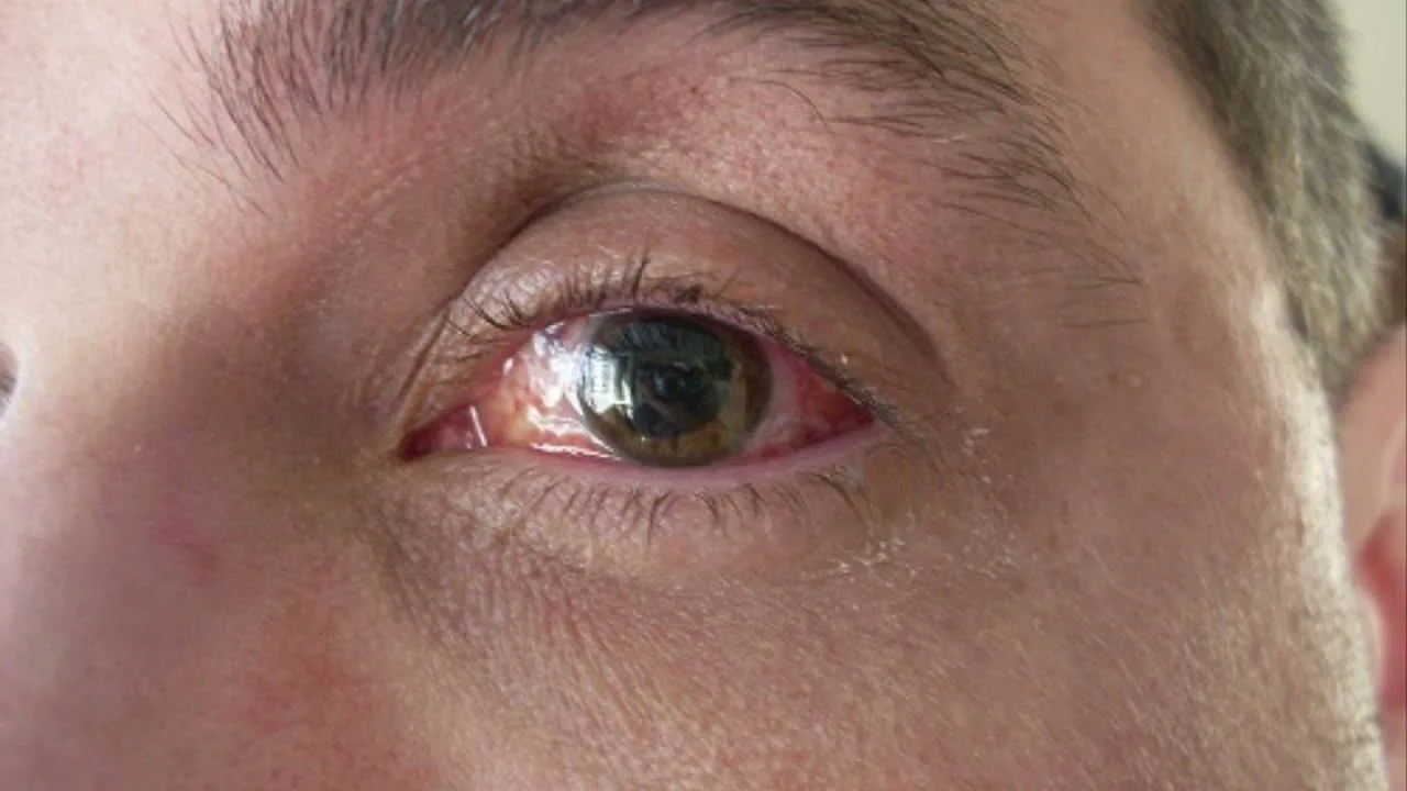
The signs of a torn retina can show themselves in many ways. They can also appear without any pain at all. Because there is no pain, people may not suspect the situation. There are things to watch for, however. If retinal tears are not treated at once they can lead to sight loss. Among the signs of a torn retina are these:
- A dark area forming in the vision
- Sight that has fallen compared with before
- Seeing floaters in the eye
- Seeing beams of light
What is the Retina?
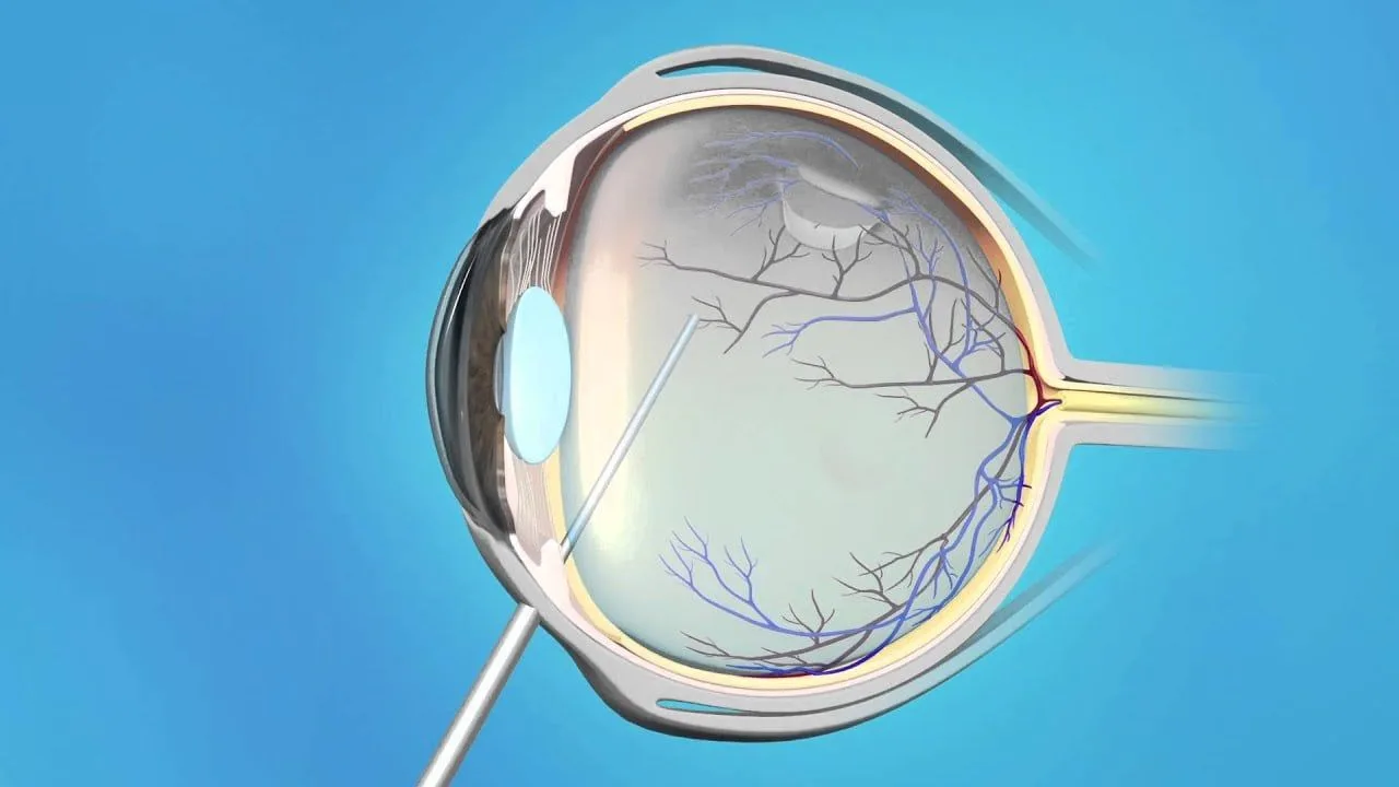
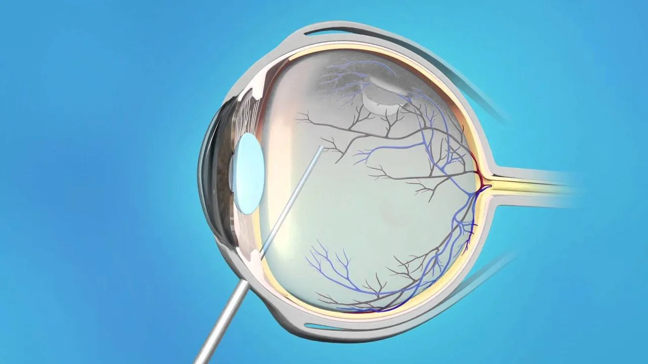
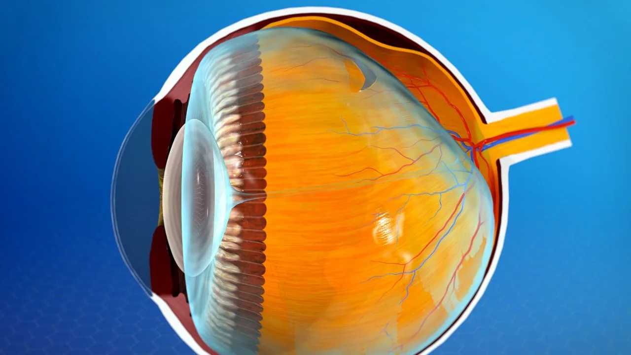
Questions such as what the retina is and what it does are among those asked about the anatomy of the eye. The retina lies at the back of the eye. This layer helps the eye perceive light and plays an important part in the process of sight. The retina, which is a layered tissue, is extremely important in terms of the ability to see. The cornea and the lens lie in the front part of the eye and carry light to the back of it. Thanks to its cells and its structure, the retina then receives that light and carries it to the brain through the optic nerves. There is a gel of about five millilitres filling the back part of the eye. It is called the vitreous gel. This gel has the consistency of egg white and sits joined to the retina. It makes up about eighty per cent of the volume of the eye. With age, the gel begins to break down over time. As the collagen fibres gather together, other fibres cause the formation of what are called pockets of water in the eye. As the collagen fibres gather and join, problems such as floaters appear in the field of vision. That shows the vitreous gel has lost its properties.A Torn Retina
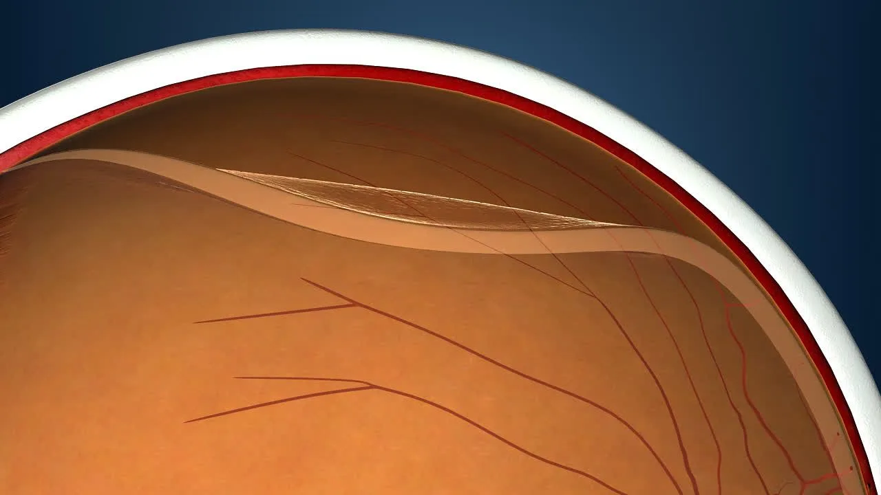
A torn retina causes floaters in the field of vision and sudden flashes of light. The situation called retinal detachment and a retinal tear are connected with one another. The blood vessels in the eye known as the choroid make up the tissue behind the eye that nourishes it. A tear in the retina can allow the retina to separate from that structure, which is called retinal detachment. If it is not treated early, permanent problems can occur in the eye. This situation leads to a reduction in vision and even to loss of the ability to see.How Does the Retina Tear?
Questions such as how the retina tears and how a retinal tear forms are of interest among people who think they are facing this situation. A retinal tear is a break in the retina. The separation of the retina from the tissue beneath it, which a tear can lead to, is called retinal detachment. The hyaluronic acid and collagen making up the vitreous gel at the back of the eye can break down over time, which causes the retina to separate from the gel. During this separation and reattachment, bursts of light are experienced in the eye. If the retina is attached to the gel abnormally, these parts pull on one another, which can lead to larger tears. If a tear is left untreated, water passes through it towards the retina, which can cause the parts in question to separate from one another again. If tears are left untreated they can lead to permanent sight loss in the long term.