




Eye Diseases
How is Retinal Vein Occlusion Treated?
- Kudret Eye Istanbul
- Last updated:
- 6 min read
- Medically reviewed by Op. Dr. Mehmet Ali Özdil
Vascular occlusion in the eye is a blockage of the blood vessels in the retinal layer at the back of the eye, the layer that allows us to see. Blockages prevent blood from reaching the retina properly and can lead to vision problems. They are grouped differently according to where the blockage lies.
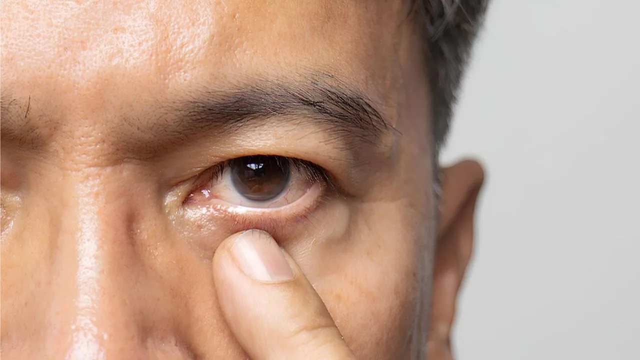
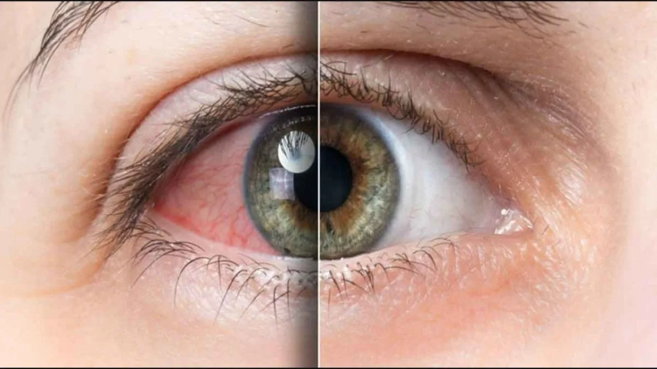
- When the main vein of the retina is blocked, central retinal vein occlusion occurs. Vision usually decreases suddenly and painlessly, and it can be linked to other conditions such as high blood pressure and diabetes.
- Hemiretinal vein occlusion is a blockage that affects half of the retina.
- In branch retinal vein occlusion, a blockage of a smaller vessel, blurred vision and black spots or thread-like shapes drifting across the eye are generally seen.
A blockage can affect not only the veins but also the artery that carries blood to the retina (retinal artery occlusion). Sudden, painless loss of vision may be caused by an artery occlusion; this is an emergency, so see an eye doctor or go to an emergency department straight away.
In people with vascular occlusion in the eye, sight loss can begin suddenly or worsen over time. Blockages in the vessels of the eye can generally be linked to chronic health problems such as high blood pressure, diabetes, high cholesterol and glaucoma. They are seen more frequently in older people in particular. If you are asking how retinal vein occlusion is treated, you can find the answer to that question and more in what follows.What Causes Vascular Occlusion in the Eye?
There are several basic causes of blockage in the vessels of the eye: Hardening of the arteries (atherosclerosis):- A build-up of fat in the arteries narrows and hardens them, which prepares the ground for blockages.
- A blood clot can block the vessels in the retina and prevent blood flow, leading to sight loss.
- Diabetes affects the vessels throughout the body and increases the risk of blockage in the eye.
- High blood pressure also hardens the vessels and can lead to clots forming.
- High pressure within the eye affects eye health by causing the retinal vessels to become blocked.
- Inflammation in the eye is among the factors that lead to vascular occlusion.
- As with every condition, smoking carries danger in eye disease too. It increases the tendency of the blood to clot and causes hardening of the vessels, raising the risk of blockage.
- High cholesterol also accelerates the formation of plaque in the vessels and can raise the pressure within them.
What Are the Symptoms of Vascular Occlusion in the Eye?
The symptoms vary with the position and the severity of the blockage. Certain signs are among the symptoms of vascular occlusion in the eye.- Partial or total sight loss can be experienced in the affected eye. This is generally regarded as the first and clearest sign of a blockage.
- Seeing black spots and lines in the field of vision is among the symptoms.
- Swelling of the macula, the area responsible for vision, can be seen.
- Abnormal new vessels forming in the eye is also among the signs.
Who Gets Vascular Occlusion in the Eye?
Vascular occlusion in the eye is seen more frequently in certain people:- Older people: the risk is higher in people over fifty in particular.
- People with cardiovascular disease: the condition appears more often in people with high blood pressure, high cholesterol and heart disease.
- Patients with diabetes: diabetes affects the health of the vessels of the eye.
- People with clotting disorders: the risk rises in those with conditions that increase the tendency of the blood to clot.
- Smokers: smoking raises the risk by damaging vascular health.
- Patients with glaucoma: where the pressure in the eye is high, blood flow can slow and lead to blockage.
- People with metabolic syndrome: this syndrome covers more than one risk factor.
How is Vascular Occlusion in the Eye Treated?
The basic aim in treating vascular occlusion in the eye is to preserve vision and halt the progression of the condition. The main treatments are:- Anti-VEGF injections: these are carried out by injecting special medicines into the eye. They are generally applied at regular intervals in order to slow the progression of the condition.
- Steroid injections: steroid medicines such as dexamethasone are injected into the eye to reduce swelling and inflammation. They are among the alternatives for patients who do not benefit from anti-VEGF treatment.
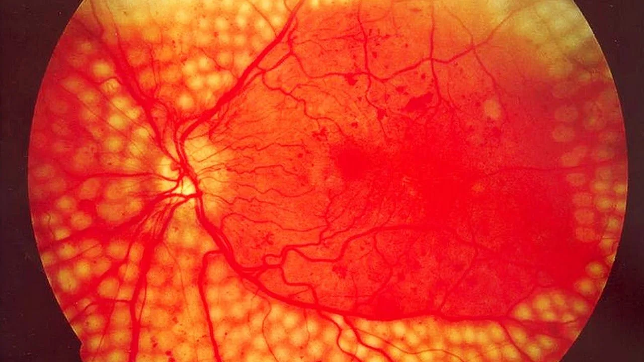
- Laser treatment (panretinal photocoagulation): this procedure is carried out to prevent new, leaking blood vessels from forming. Small laser burns are created in the retina during the procedure.
Treatment of Vascular Occlusion in the Eye
Vascular occlusion in the eye is treated with different methods according to the patient's situation and the severity of the condition. The aim of treatment is to preserve vision and prevent possible complications.Intraocular Injections
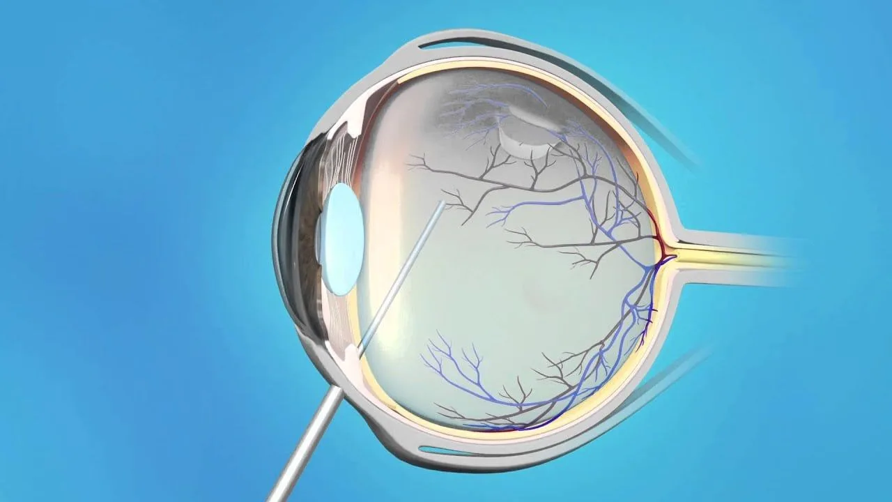
This method is generally the treatment used most often. After dilating the pupils and numbing the eye with local anaesthesia, the doctor injects the medicine into the eye with a fine needle. These medicines aim to prevent swelling over the retina and the formation of new vessels. The procedure is short, and the eye is cleaned afterwards.Laser Treatment
Laser treatment is used above all to prevent abnormal blood vessels from growing. After the eye has been numbed and a contact lens fitted, the doctor makes small laser applications to the retina, halting the development of those abnormal vessels.Surgical Treatments
In certain particular situations surgery may be needed. One such operation is pars plana vitrectomy. During it the gel within the eye, the vitreous, is removed so that the pressure on the retina is reduced and bleeding cleared. This operation is generally chosen in patients who do not respond to other treatment. The course of treatment is planned according to each patient's needs.Frequently Asked Questions
- 1
Does vascular occlusion in the eye heal completely?
The blocked vessel generally cannot be reopened, but with treatment vision can be preserved and complications prevented. - 2
What are the symptoms?
Usually painless sight loss that develops suddenly or slowly, black spots in the field of vision and blurred vision. - 3
Who is at risk?
People over fifty, and those with high blood pressure, diabetes, high cholesterol or glaucoma, and smokers. - 4
What is the aim of treatment?
To preserve vision, reduce macular oedema, prevent new vessels from forming and prevent complications. - 5
Can it be prevented?
Yes. Regular eye examinations, control of blood pressure, blood sugar and cholesterol, giving up smoking and a healthy life all reduce the risk. What is Hyperopic Astigmatism?