




Eye Diseases
What is the Eye and What is Its Anatomy?
- Kudret Eye Istanbul
- Last updated:
- 7 min read
- Medically reviewed by Op. Dr. Mehmet Ali Özdil
The answer to what the eye is and what it is like matters a great deal for eye health. The eye is one of the most important senses, allowing living creatures to see and to perceive this world. It carries very great importance in how we perceive our surroundings. Knowing the anatomy of the eye makes it far easier to solve many of the problems and conditions relating to it. Problems affecting the eye need to be identified and the appropriate treatment applied. The eye forms the main part of a highly complex system that makes sight possible.
The eye consists of three sections in all. They are:
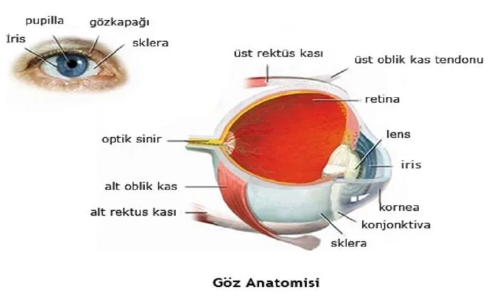
- The outer layer, holding the cornea and the sclera
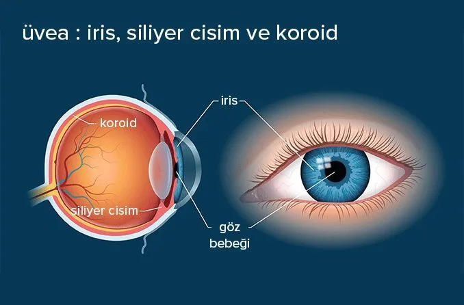
- The middle (vascular or uveal) layer, holding the iris, the ciliary body and the choroid
- The inner layer, holding the retina
The Benefits of Knowing Eye Anatomy
The benefits of knowing eye anatomy are extremely important in medicine. Understanding the structure of the eye matters for treating glaucoma, cataract and other vision problems. Among the most common vision problems today are myopia, hyperopia and astigmatism. The reason for difficulty seeing at distance, near and intermediate ranges is usually the length of the eyeball or the curvature of the cornea. If the anatomy of the eye were not understood, reaching solutions to these problems would be very difficult. In the same way, fitting intraocular lenses, treating cataract and treating other vision problems have all emerged through knowledge of eye anatomy. Because that anatomy is known, which part a problem arises from is easily understood. Laser surgery is carried out straightforwardly today for the same reason. Although the eye consists of many parts, it in fact represents a whole, and understanding the parts of that whole matters in identifying the problems arising within it.
The Structure of the Eye
The question of what the structure of the eye is like is among the subjects most widely asked about. The eye works by perceiving the light around humans and other living creatures and carrying images to the brain. Although the anatomy of the eye is complex, it is extremely important. Light and images form through the optic nerve system and its connection to the other nerves. That light is refracted and carried to the back of the eye, and from there it is carried to the brain through the nerves so that the function of sight takes place. Many structures are involved in the anatomy of the eye. They are important, and if they are not healthy serious vision problems can follow.The Cornea
The cornea is the transparent structure forming the outer layer. It has a curvature of its own and is positioned over the structures in the middle section such as the lens and the iris. It is like a protective layer at the front of the eye. The cornea consists of many layers. A healthy cornea allows light to be refracted correctly, which provides sharp and clear vision. It is the first place light reaches in the process of sight. The cornea is transparent because it contains no blood vessels. The absence of blood vessels in this part keeps the cornea transparent. With its vessel-free and transparent structure, the cornea allows light to pass unobstructed to the layers within the eye. Thanks to nerves sensitive to external stimuli, the blink reflex is triggered. The clear watery fluid (aqueous humour) filling the anterior chamber between the cornea and the iris, and the posterior chamber between the iris and the lens, is produced in the ciliary body and drains through the canal of Schlemm, balancing the pressure within the eye. If that flow is disturbed, glaucoma can develop.
The Pupil and the Iris
The iris is the coloured part of our eye. It lies around the pupil. The pupil widens and narrows according to the amount of light, and this happens through the iris. The pupil determines the amount of light passing through it. When a light is shone into your eye, for instance, the pupils narrow and less light passes through, which provides a clearer image in bright light. In dim light, however, the pupils widen so that more can be seen and more light gathered. The pupil and the iris move together so that light is carried into the eye in a controlled way. The ciliary muscle, located in the ciliary body behind the iris, changes the curvature of the lens by relaxing and tightening the zonular fibres. This mechanism allows us to focus clearly at near and at distance, which is known as accommodation.The Lens
The lens is among the most important parts that come to mind when eye anatomy is mentioned. It is positioned just behind the iris and in front of the vitreous body. It is an important structure lying between the iris and the vitreous body. It is transparent and allows a sharp and clear image to form. The gel-like vitreous humour filling the space between the lens and the retina preserves the shape of the eyeball and supports the retina from behind. As it liquefies with age it can cause floaters.The Retina
The retina lies at the back of the eye. It is one of the main parts of the function of sight that takes place as light enters the eye. Light projected by the lens is carried to the cells in the retina. The retina is a multi-layered structure made up of photoreceptors (rods and cones), interneurons and ganglion cells. It allows detail to be perceived more easily so that good vision is provided. The macula and fovea at the very centre of the retina are the area where cone photoreceptors are concentrated, and they provide colour vision and high resolution. The optic disc, the area of the retina containing no photoreceptors, is the point where the nerve fibres leave; the image falling there is not perceived as a blind spot in the brain.The Optic Nerves
A single optic nerve leaves each eye. It carries the visual information, converted into electrical signals in the retina, to the brain. The optic nerve leaves the posterior pole of the eye at the optic disc and extends towards the visual centre of the brain. The optic nerves lie at the back in the anatomy of the eye. Beginning at the back of the eye, they reach the visual part of the brain and allow visual signals to be carried. Beyond these parts, the other parts of the eye are:- Sclera: the white part of the eye. It protects the eye and is firm and durable.
- Eye muscles: the muscles that allow the eyes to move.
- Tear glands: the eyes are structures that must stay moist at all times, and the tear glands keep them so. When a foreign body enters the eye they actively make it easier to be rid of it. The tear film consists of three layers: the oil layer from the meibomian glands limits evaporation, the watery layer cleans the cornea, and the mucin layer spreads the tears evenly over the surface of the cornea.
- Conjunctiva: the thin mucous membrane covering the inner surface of the eyelids and the front surface of the sclera, which moistens the eye. It does not cover the cornea. It is the inner part of the eyelid that keeps the eye moist and protected.
- Eyelids: they protect the eye through the lashes and the skin.