Our eye health is among the most delicate subjects we neglect without noticing amid the intensity of life. When people speak of retinal thinning, what is generally meant is peripheral retinal thinning (lattice and similar degenerations) or retinal changes caused by high myopia. These situations often give no symptom and are noticed during a routine examination of the back of the eye. The retina is one of the most important structures of the eye. Thinning occurring in the retina causes no problems at all in most people; in some, however, it can pave the way over time for a retinal tear and a retinal detachment. Recognising the warning signs of a tear or detachment therefore matters a great deal in preserving sight.
What is Retinal Thinning?
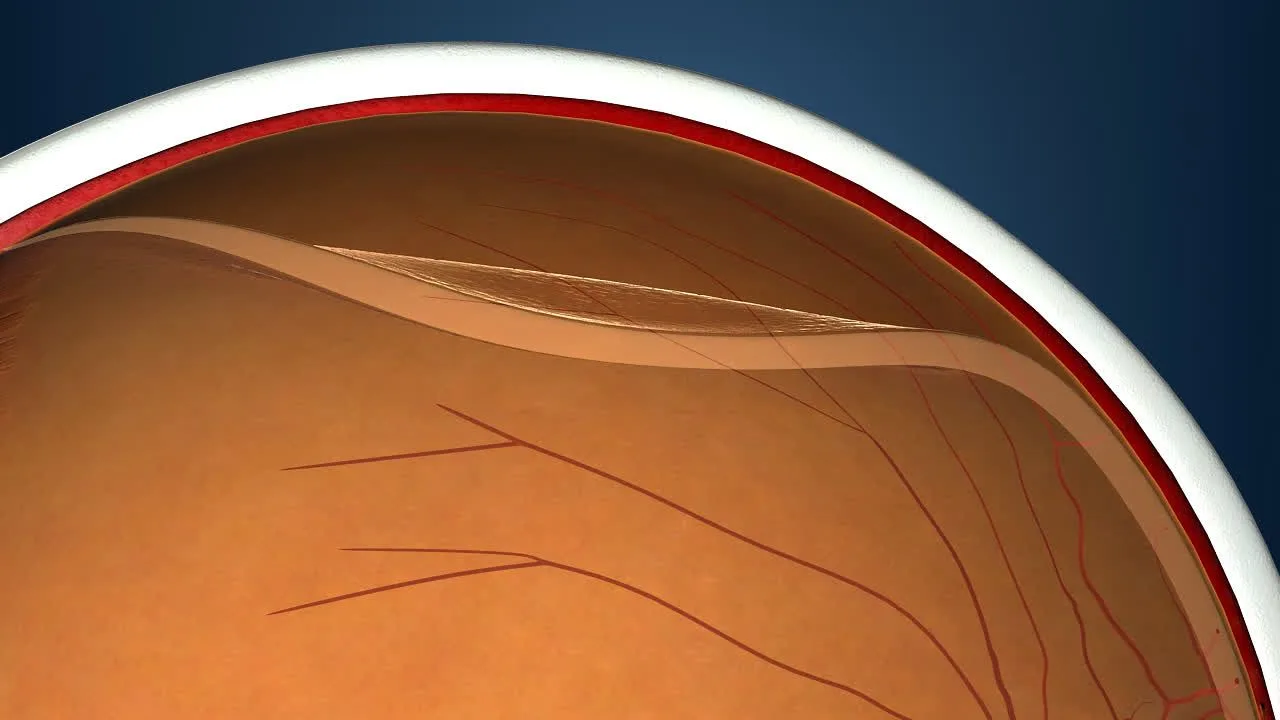
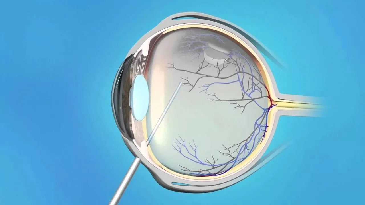
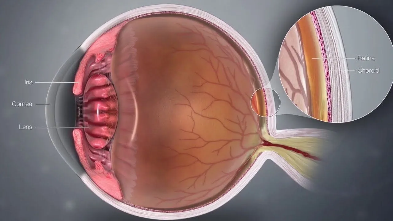
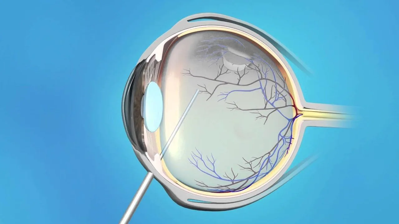
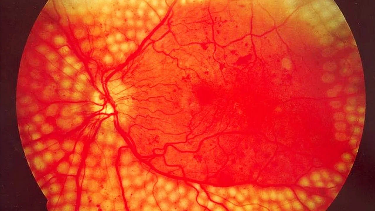
The retina is the tissue at the back of the eye that perceives light, turns it into nerve signals and carries them to the brain. In lattice degeneration there are thinned, weaker areas in the outer (peripheral) part of the retina. The gel that fills the eye (the vitreous) can be more firmly attached at the edges of these areas. If the vitreous pulls on these points as it separates from the retina with age, a retinal tear can form. The thinning itself usually does not affect vision.
Retinal thinning
What Are the Symptoms of Retinal Thinning?
Retinal thinning usually causes no symptoms and is found during a routine examination of the back of the eye. What are called the symptoms of retinal thinning are really the warning signs of a tear or detachment that may develop in the thinned area. If any of the following occurs, see an ophthalmologist without delay; early consultation can be critical in stopping a tear from progressing to a detachment:
People with retinal thinning meet black spots, thread-like shapes or images resembling a spider's web moving around within the eye. Floaters can normally be seen in everyone. A sudden increase in floaters, particularly together with flashes of light, can indicate that a retinal tear has developed and calls for an examination the same day.
Flashes of light
One of the strongest and most important warnings of retinal thinning is flashes of light. If you see small bursts like lightning as the eye moves, as you enter a dark room or as you suddenly shift your gaze, that suggests a risk of traction or a tear on the retina. It is distinguished through a dilated examination of the back of the eye. As the traction increases, the flashes of light increase as well.
The formation of a shadow
If a tear develops and progresses to a retinal detachment, a dark shadow appears at the corner of the field of vision. The shadow generally does not stay still; it grows and widens from below upwards or horizontally. A curtain or dark shadow across vision, and a sudden reduction in sight, can be warnings of an emergency such as retinal detachment, where the retina separates from its place, and rapid assessment should be recommended. If you notice the shadow growing, that means a certain part of the retina has begun to lose its function.
Straight lines looking wavy, colours fading or central vision gradually declining are not symptoms of retinal thinning; they are usually signs of conditions affecting the macula (the centre of the retina). These complaints also call for an eye examination.
How Does Retinal Thinning Form?
Retinal thinning does not form all at once. What sets the process off is generally the structural changes the eye undergoes over the years. Among the most common causes are:
Advanced myopia
The gel within the eye changing with age
Injuries and blows
Genetic predisposition
Having had a retinal problem before
Chronic conditions such as diabetes
In people with high myopia the eyeball lengthens, so the retina naturally thins and becomes more prone to tearing. In some people retinal thinning can lead to a retinal tear and then to retinal detachment. Detachment means the retina has separated from its place. The situation calls for urgent intervention. If that intervention is delayed, the person experiences permanent sight loss.
Anyone experiencing the warning signs should see an ophthalmologist without losing time. An examination of the back of the eye with the pupil dilated shows where the thinning is and whether there is a tear, and the decision between monitoring and treatment is made accordingly.
Treatment of Retinal Thinning
Most retinal thinning needs no treatment; the aim is to prevent a tear or detachment, or to catch it early. The treatment process is shaped by the degree of the thinning, by the person's risk factors and by whether there is a tear in the retina. Early diagnosis affects the success of the treatment applied directly and helps the person preserve their capacity to see.
The approach is as follows:
Monitoring
Thinning that causes no symptoms and contains no tear usually needs no treatment. The back of the eye is examined at the intervals your doctor recommends, and the warning signs are explained to you.
Prophylactic laser where needed
If a tear or hole is found in the thinned area, or if the person's risk is high (for example after a detachment in the other eye), the doctor may recommend prophylactic laser (laser photocoagulation). The laser bonds the retina around the tear or thinned area, aiming to reduce the risk of a detachment. Not every case of retinal thinning needs laser.
Surgery if a detachment occurs
If a retinal detachment develops, laser alone is not enough and surgery is needed. A detachment is urgent; the sooner it is operated on, the better the chance of preserving sight.
Healthy Living with Retinal Thinning
Alongside medical treatment, certain adjustments to lifestyle help protect the retina as well.
Doctors make the following recommendations:
A diet that keeps blood sugar in balance
Regular eye examinations
Protecting the eyes from injury
Reducing the consumption of tobacco and alcohol
Increasing foods rich in omega 3
Paying attention to the points above keeps the risks associated with retinal thinning to a minimum and provides protection.
Frequently Asked Questions
1
Does retinal thinning resolve by itself?
Most areas of peripheral degeneration do not simply disappear. What matters is regular follow-up and timely intervention where there are findings that carry risk. It varies from person to person, so an assessment by an ophthalmologist is needed.
2
Do floaters always mean a retinal tear?
No. Floaters can be seen commonly. If a sudden increase, flashes of light or a curtain across vision accompany them, however, an examination the same day is recommended.
3
Is the risk greater in people with high myopia?
Yes, high myopia can increase the risk of certain peripheral retinal lesions and of a retinal tear or detachment. Regular examination of the back of the eye therefore matters.
4
Is prophylactic laser applied to every case of retinal thinning?
Generally no. The decision to use laser is taken according to the presence of a tear or hole, the type of the lesion and personal risk factors. It is not compulsory for every patient.
5
What are the symptoms of retinal detachment and when is it an emergency?
A curtain or dark shadow, a sudden reduction in vision and a sudden increase in floaters together with flashes of light are urgent warnings. An ophthalmologist should be consulted without delay.
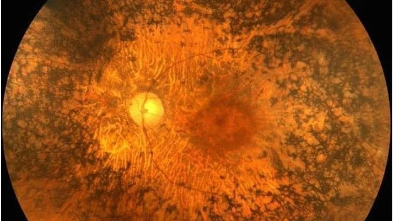
Retinitis pigmentosa is one of the causes of night blindness, although the two are not the same thing. It is one of the genetic eye conditions. In the early stages people generally consult a doctor saying their vision at night has fallen and that they have difficulty seeing.
A retinal tear is a break in the retinal layer of the eye that can lead to the retina separating from the place it is attached to. It is a very serious eye condition. Its symptoms are a sudden increase in floaters, flashes of light and darkening of vision.
Vascular occlusion in the eye is a blockage of the blood vessels in the retinal layer at the back of the eye, the layer that allows us to see. Blockages prevent blood from reaching the retina properly and can lead to vision problems.
Diabetic retinopathy is the greatest cause of sight loss arising from diabetes. Raised blood sugar damages the blood vessels, and diabetes affects the nerves of the eye and the capillaries within the nerve layer.
Essential cookies are always on. With your permission we also use Google and Meta cookies for measurement and ads.Cookies needed for the site to work are always on. With your permission we also use Google and Meta cookies to measure visits and show you relevant ads. You can change your choice at any time with the "Cookie settings" link at the bottom of the page.Detailed Information