




Eye Diseases
What is Eye Angiography?
- Kudret Eye Istanbul
- Last updated:
- 5 min read
- Medically reviewed by Op. Dr. Mehmet Ali Özdil
The questions of what eye angiography is and how it is carried out are of considerable interest to those meeting the method for the first time. New developments occur in eye health day by day. These technologies have reached such an advanced level that even intervening in a structure as delicate and complex as the eye has become straightforward.
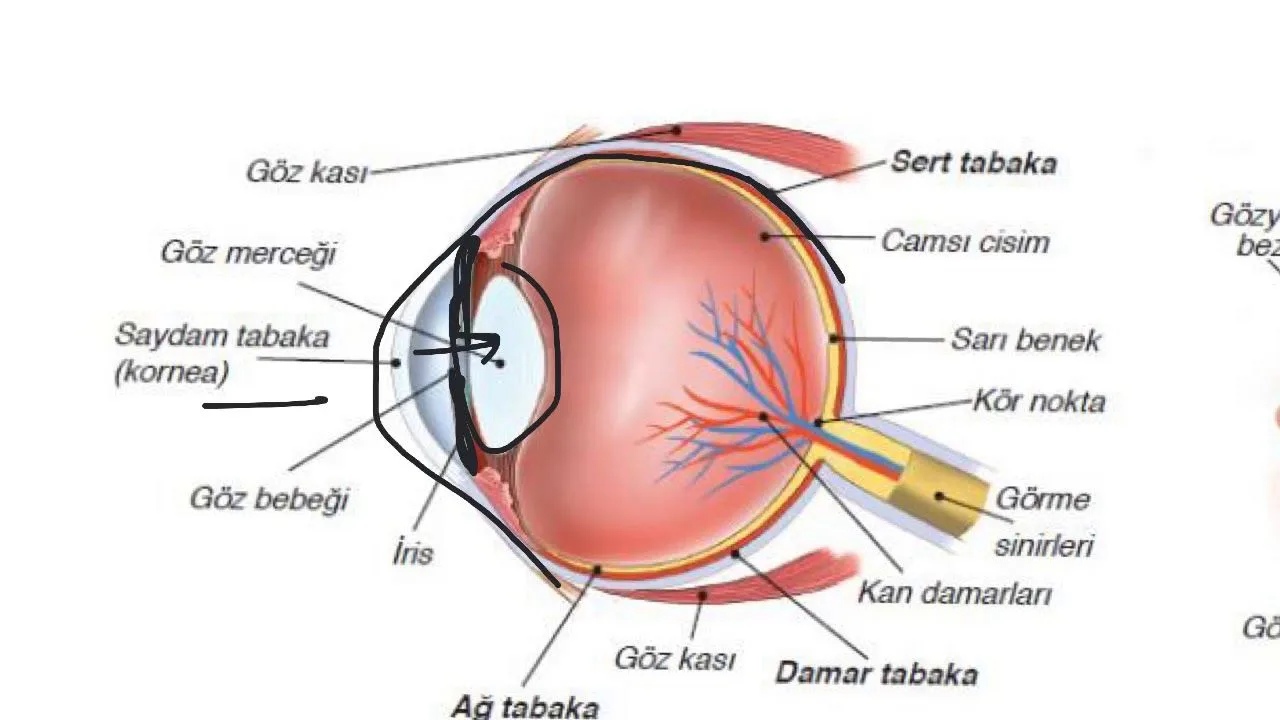
Anatomically the eye consists of a front, a middle and a back section. Eye angiography is essentially the photographing of the retinal and choroidal vessels after they have been stained, using fluorescein fundus angiography (FFA) or indocyanine green angiography (ICG). Circulation disorders in the posterior segment of the eye are examined in detail this way. Through the images obtained, the following conditions can be identified:
- Diabetic retinopathy
- Age-related macular degeneration
- Retinal vein and artery occlusion
- Central serous chorioretinopathy
- Tumours of the eye
- Uveitis
- Conditions of the retina
How is Eye Angiography Carried Out?
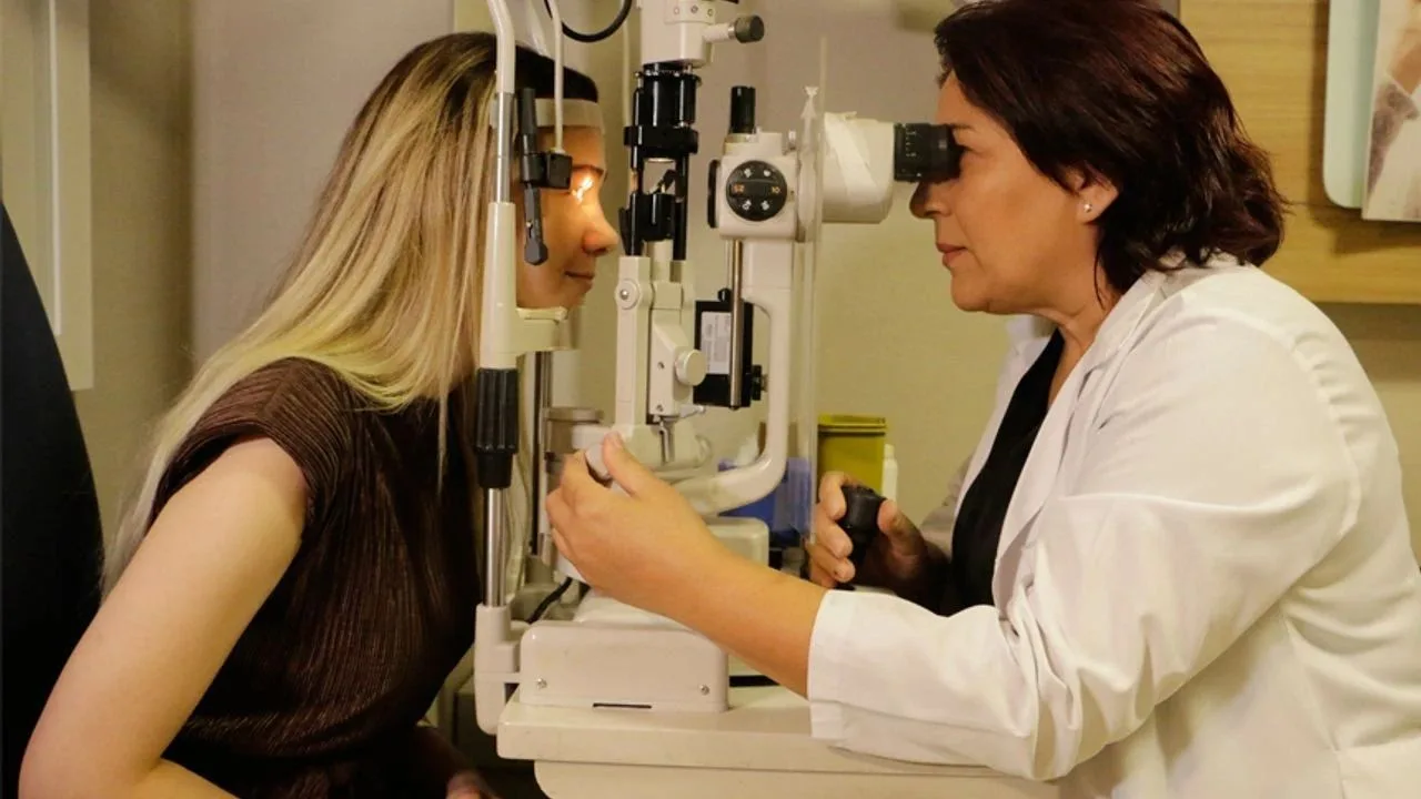
The questions of how eye angiography is carried out and whether it is risky are of interest to patients. Patients feel no pain during the procedure. A special drop is applied to dilate the pupil. This is painless and takes a short time. The dilated pupils are then examined with a special machine. Light is used to photograph the area at the back of the person's eye. Thanks to the drops applied and the substances given, the vessels at the back become much more visible. The leaks, breaks and retinal problems in those visible vessels are identified easily this way. Through eye angiography, defects of the eye are seen very clearly. This method, which gives very clear results, carries very little risk. No pain, soreness or discomfort is felt during eye angiography.How Long Does Eye Angiography Take?
Eye angiography takes a very short time. The vessels at the back of the eye are stained with special substances and dyes, so that problems which would otherwise be invisible are revealed. It is among the most reliable methods for reaching a diagnosis. Before eye angiography, special eye drops are applied to the patient. No needle, cutting instrument or anything else that enters the eye is used in this procedure. The machine used in eye angiography allows the back of the eye to be imaged through the pupil. No pain or soreness occurs during the procedure. The light and lens used make it easier to see inside the eye. Drops are used to dilate the pupil and fluorescein to make the vessels change colour and stand out. The symptoms met most often after the procedure are nausea and a change in the colour of urine. Both the nausea and the coloured urine can come from the fluorescein dye. Because no pain is felt, this straightforward technique is used in a great many situations. The fluorescein given into the veins makes the vessels of the eye stand out, so that a clear map of the vascular system emerges. Breaks, blockages and leaks in the vessels can be imaged easily this way. The procedure takes approximately ten to twenty minutes.
When is Eye Angiography Carried Out?
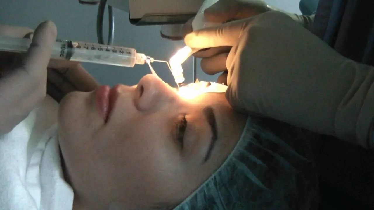
Another question asked about this method is when eye angiography is carried out. A great many methods are used to image the eye when eye health is assessed. Methods such as measuring eye pressure are carried out with machines, and patients bring their eyes close to them. Through the light and lenses in the machine, the inside of the eye is imaged. In eye angiography too, drops are first applied to dilate the pupils, so that the inside of the eye can be imaged through them. A line is opened in the arm and a special substance is given so that the vessels within the eye take on colour. The procedure is very short. There are many reasons for carrying out eye angiography. They are:- People with a tumour or inflammation of the eye (uveitis)
- Patients with diabetes
- Macular oedema caused by diabetes or retinal vein occlusion
- Identifying vascular occlusion in the eye
- Central serous chorioretinopathy (fluid build-up in the eye)
- Identifying and treating age-related macular degeneration
- Identifying bleeding in the eye