




What Causes a Tear in the Eye? Causes of a Retinal Tear
- Kudret Eye Istanbul
- Last updated:
- 8 min read
- Medically reviewed by Op. Dr. Mehmet Ali Özdil
A tear inside the eye is most often a tear in the retina, the layer that lines the back wall of the eye (a retinal tear). The most common cause is the vitreous, the clear gel that fills the eye, shrinking with age and pulling on the retina as it separates from it. High myopia, a blow to the eye, a family history of retinal tears and previous eye surgery all increase the risk.
What Is a Retinal Tear?
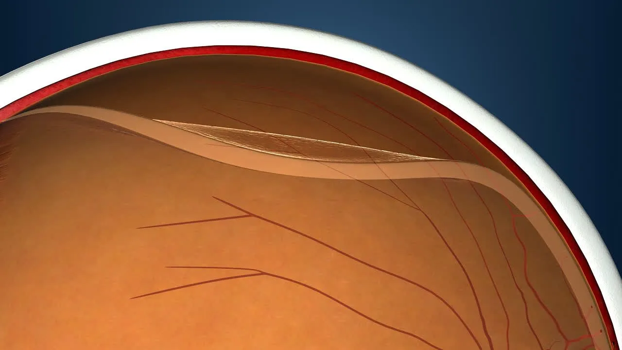
A retinal tear is a break in the light-sensitive retina that can progress to a retinal detachment if it is not treated. Tears can form in one or more places in the retina, most often near its outer edges.
If fluid seeps through the tear and under the retina, the retina can lift away from its normal position; this is called a retinal detachment. If a tear is caught before a detachment develops, it can be sealed with a laser; once a detachment has developed, the treatment is surgery.
What Causes a Tear in the Eye?
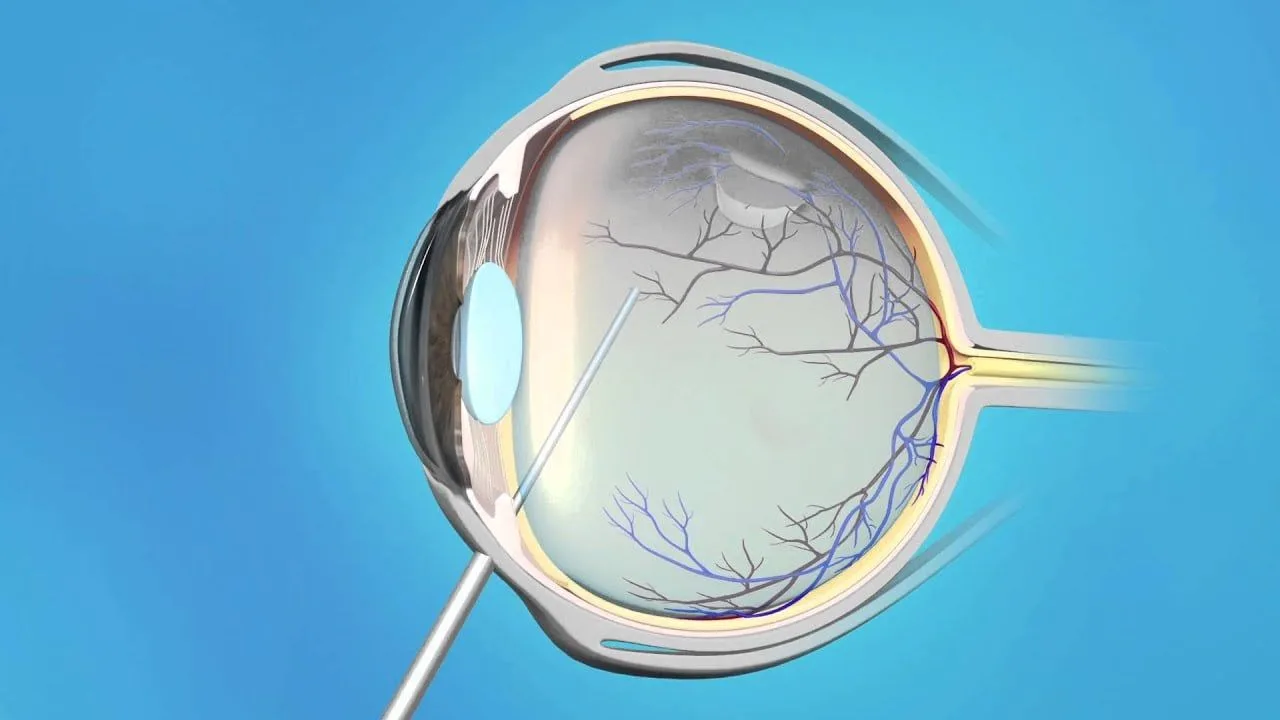
The most common cause is the vitreous shrinking with age and pulling on the retina as it comes away. In young people, the vitreous is a firm, clear gel. Over the years it becomes more liquid, shrinks and starts to pull away from the retina.
This age-related separation of the vitreous from the retina is called a posterior vitreous detachment. It becomes more common especially after the age of 50 and causes no problems for most people. However, if the vitreous pulls on the retina at a point where the two are firmly attached, a tear can form.
A sudden increase in floaters and the appearance of flashes of light can be a sign that the vitreous has started to pull on the retina.
What Increases the Risk of a Retinal Tear?
The main risk factors are age, high myopia, a blow to the eye, a family history of retinal tears and previous eye surgery.
| Risk factor | Why it increases the risk |
|---|---|
| Age | The vitreous shrinks with age and can pull on the retina as it separates |
| High myopia | The eyeball is longer, so the retina is thinner |
| A blow to the eye | A sudden impact can cause a tear in weaker areas of the retina |
| Family history of retinal tears or detachment | Increases the risk |
| Previous eye surgery (e.g. cataract surgery) | The vitreous may separate sooner after surgery inside the eye |
| Naturally thin areas in the retina | Can be seen during an examination; preventive laser may be considered if needed |
| A previous retinal tear in one eye | The risk of a new tear in the same or the other eye may be higher |
High Myopia
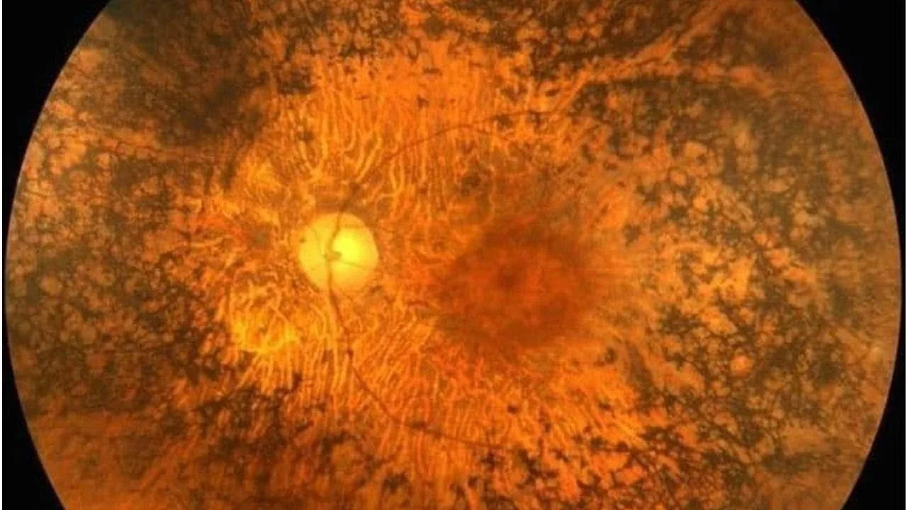
In high myopia (strong nearsightedness), the eyeball is longer than normal, which makes the retina thinner. A thinner retina tears more easily when the vitreous pulls on it. High myopia is generally defined as -6 or more, although the risk can also be slightly higher than normal at lower prescriptions. If you have high myopia, regular retinal check-ups are recommended.
A Blow to the Eye
An impact during sport or an unexpected accident suddenly changes the shape of the eyeball and the pressure inside it. This sudden change can cause a tear in weaker areas of the retina. Blows taken in contact sports such as boxing, football and basketball carry this risk too.
Family History of Retinal Tears
A family history of retinal tears or retinal detachment increases the risk. In this case, regular eye examinations are recommended.
Previous Eye Surgery and Earlier Tears
The risk of a retinal tear can be higher after surgery inside the eye, such as cataract surgery. This risk is low, but if you notice floaters or flashes of light after surgery, it is important to let your doctor know. People who have had a retinal tear in one eye are also more likely to develop a new tear in the same or the other eye.
Diabetes and Other Eye Conditions
In advanced diabetic retinopathy, new blood vessels and scar tissue that form on the retina can pull on it and cause it to detach. Some degenerative conditions affecting the retina can also weaken it and raise the risk of a tear.
Long-lasting inflammation inside the eye (uveitis) can change the structure of the vitreous and increase the pulling force where the vitreous is attached to the retina. In some people, this can raise the risk of a retinal tear.
Naturally Thin Areas in the Retina
Some people are born with thin, weaker areas near the outer edges of the retina. These areas can tear even with slight pulling from the vitreous. They can be seen during an examination with dilated pupils, and your doctor may recommend monitoring or, if needed, preventive laser treatment. You can read more in our article on retinal thinning.
What Are the Symptoms of a Retinal Tear?
A retinal tear can sometimes form without any symptoms, but it often shows itself through warning signs such as a sudden increase in floaters and flashes of light.
- A sudden increase in floaters
- Flashes of light starting or becoming more frequent
- A shadow in part of your field of vision
- Sudden blurry vision
- Darkening, as if a curtain is coming down over your vision
You can find more detail in our article on the symptoms of a retinal tear.
When Should You See an Eye Doctor Straight Away?
If you suddenly notice many new floaters, if flashes of light start or increase, or if a shadow like a curtain falls over part of your vision, see an eye doctor the same day. These symptoms do not always mean a tear, but catching a tear before a detachment develops makes treatment easier.
How Is a Retinal Tear Diagnosed?
A retinal tear is diagnosed with a retinal examination after your pupils have been dilated with drops. If there is bleeding inside the eye and the retina cannot be seen clearly, your doctor may also order an ultrasound scan of the eye.
If you have high myopia, a family history of retinal disease or have had eye surgery, your eye doctor may check your retina with dilated pupils. Wearing protective eyewear during contact sports lowers the risk of injury.
How Is a Retinal Tear Treated?
If a retinal tear is caught before a detachment develops, it can be sealed with a laser. The laser creates tiny scar points around the tear; as they heal, they bond the retina to the tissue underneath and stop the tear from spreading. The procedure is usually done with anaesthetic eye drops and without a hospital stay.
If laser treatment is not possible, for example because of where the tear is or because of bleeding inside the eye, freezing treatment applied from outside the eye (cryotherapy) may be used. An untreated tear can progress to a retinal detachment; once a detachment has developed, the treatment is surgery.
How much vision returns after retinal detachment surgery depends on how widespread the detachment is and whether the macula, the centre of the retina, was affected. Your doctor will decide on the right treatment after examining you; you can find the options on our retina treatments page.
Frequently Asked Questions
- 1
Can a retinal tear heal on its own?
Retinal tears that cause symptoms generally do not close on their own and usually need treatment. Some small, older tears that cause no symptoms may only need monitoring. Your eye doctor will decide whether treatment is needed based on the type and location of the tear.
- 2
What is the difference between a retinal tear and a retinal detachment?
A retinal tear is a break in the retina. A retinal detachment is when the retina lifts away from its normal position; an untreated tear can progress to a detachment. If a tear is caught before a detachment develops, it can be sealed with a laser; once a detachment has developed, the treatment is surgery.
- 3
Are retinal tears more common in people with myopia?
In high myopia, the eyeball is longer and the retina thinner, so the risk of a retinal tear is higher. If you have high myopia, regular retinal check-ups are recommended.
- 4
Is a retinal tear painful?
No, a retinal tear is usually painless because the retina has no pain-sensing nerve endings. The warning signs are not pain but a sudden increase in floaters, flashes of light and a shadow in your field of vision.
- 5
How long does laser treatment for a retinal tear take?
Depending on the size of the tear, the laser procedure usually takes from a few minutes to 15-20 minutes and is done with anaesthetic eye drops. Most people return to daily life the next day, and full healing, when the laser scars firmly bond the retina, takes about 2 weeks. You may be asked to avoid heavy lifting and strenuous sport during this time.
- 6
If one eye has a retinal tear, will the other eye get one too?
If you have had a tear in one eye, the risk of a tear in the other eye is higher than normal. For this reason, it is recommended that the other eye is also examined with dilated pupils.
- 7
Can high eye pressure cause a retinal tear?
High eye pressure (glaucoma) is not one of the known main causes of retinal tears; glaucoma mainly affects the optic nerve. As both conditions can occur in the same person, regular eye examinations are still important.